THE BIOLOGY PRIMER · LAB
The Origin and Evolution of Animals
Lab Overview
Use the illustrations on this page as visual guides to complete the required drawings on paper. Your drawings do not need to match the illustrations exactly. Focus on the overall form and the important structures. Drawing requires you to actively identify shapes, relationships, and labels instead of simply looking at them, which helps you learn and remember the organisms more effectively. Drawings are evaluated for completion, not artistic quality. The phylogeny exercises are evaluated for accuracy.
The phylogeny exercises require you to apply what you learn throughout the lab. Read the lab text carefully and use the organisms, structures, and evolutionary traits you encounter to determine where each characteristic belongs on the phylogenies. The completed phylogenies are not provided on this webpage.
Learning Goals
- Recognize major animal groups and the body features used to distinguish them.
- Connect animal body plans and developmental traits with evolutionary relationships.
- Use morphological characteristics as synapomorphies when interpreting an animal phylogeny.
Jump to Exercise
Exercise A: The Origin of Animals
Phylum Choanozoa (Choanoflagellates)
Origin of Animals Phylogeny
A synapomorphy is a shared characteristic that helps identify a branch of a phylogeny. Use the descriptions in your printed lab to compare Fungi, Choanozoa, and Animalia. For each branch, identify which group or groups descend from it, then choose the characteristic from the word bank that matches all of those descendants.
Fill all five boxes before checking your answers.
Exercise B: Porifera
Phylum Porifera (Sponges)
Exercise C: Diploblastic Animals
Phylum Cnidaria (Sea Anemones, Coral, Jellyfish, Hydras)
Phylum Ctenophora (Comb Jellies)
The printed lab introduces Cnidaria and Ctenophora together as animals with a primarily two-layered developmental organization. Current evidence treats Cnidaria and Ctenophora as separate evolutionary lineages, and their developmental biology is not identical.
For this exercise, focus on comparing their body forms, symmetry, feeding structures, and nervous organization while using the terminology and sequence provided in your printed lab.
Exercise D: Triploblastic Animals
Characteristics of Triploblasts
The printed lab calls this stage a “triploblastic blastula.” More precisely, the three germ layers become established during gastrulation. The drawing guide therefore shows a gastrula with ectoderm, mesoderm, and endoderm.
Exercise E: Acoelomorpha
Phylum Acoelomorpha (Acoelomorphs)
Modern biology avoids describing Acoelomorpha as “primitive.” Acoelomorphs are an early-diverging bilaterian lineage with their own evolutionary history, and their exact position near the base of Bilateria continues to be studied.
Exercise F: Coelomates (Protostomes and Deuterostomes)
Protostome and Deuterostome Development
Blastopore fate and coelom formation are useful classical comparisons between protostomes and deuterostomes, but they are not universal rules. Development varies among lineages, and modern classification also relies heavily on molecular evidence.
Exercise G: Protostomes Overview
Lophotrochozoa
Ecdysozoa
Incremental skeletal growth does not characterize all lophotrochozoans. Treat the clam here as an example of shell growth. In contrast, molting, or ecdysis, is a defining feature of Ecdysozoa.
Exercise H: Protostomes – Lophotrochozoa
Phylum Bryozoa (Moss Animals)
Phylum Rotifera (Rotifers)
Modern phylogenies place these animals within the broader protostome clade Spiralia. Rotifers are generally placed within Gnathifera rather than Lophotrochozoa in the narrower sense. Continue using the printed lab sequence for this exercise.
Phylum Platyhelminthes (Flatworms)
Phylum Annelida (Segmented Worms)
Phylum Mollusca (Mollusks)
Exercise I: Protostomes – Ecdysozoa
Phylum Nematoda (Roundworms)
Phylum Tardigrada (Waterbears)
Phylum Onychophora (Velvet Worms)
Phylum Arthropoda (Arthropods)
Subphylum Myriapoda: Class Chilopoda (Centipedes)
Subphylum Hexapoda: Class Insecta (Insects)
Subphylum Chelicerata (Chelicerates: Spiders, Ticks, Mites, Horseshoe Crabs)
Subphylum Crustacea (Crustaceans: Lobsters, Crabs, Shrimp)
Exercise J: Deuterostomes Overview
Acorn worms are not an evolutionary “link” between invertebrates and vertebrates. Hemichordates are an independent lineage grouped with echinoderms in Ambulacraria. They share some features with chordates but are not intermediate chordates.
Exercise K: Deuterostomes - Echinodermata
Class Asteroidea (Sea Stars)
Class Ophiuroidea (Brittle Stars)
Class Echinoidea (Urchins, Sand Dollars)
Exercise L: Deuterostomes - Chordata
Subphylum Cephalochordata (Lancelets)
Subphylum Agnatha - now Class Hyperoartia (Lampreys)
Class Chondrichthyes (Sharks, Rays, Skates)
Class Actinopterygii (Ray-finned Fish)
Class Sarcopterygii (Lobed-finned Fish)
Class Amphibia (Amphibians: Frogs, Salamanders, Newts, Caecilians)
Class Reptilia (Reptiles)
Class Mammalia (Mammals)
Living vertebrate groups are branches of a shared evolutionary tree, not stages of progress. Use traits such as jaws, limbs, amniotic eggs, hair, and mammary glands to identify branching relationships rather than levels of evolutionary advancement.
Exercise M: Animal Phylogenetics
Place each evolutionary characteristic into the correct numbered box on the phylogeny. Select a phrase and then select a box. On a desktop computer, you may also drag phrases directly to boxes. Use Check My Answers whenever you want feedback.
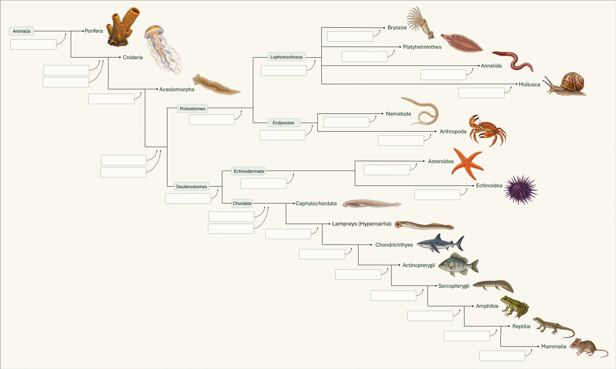
Tip: Select a phrase, then select any unlocked numbered box. On desktop, you can also drag a phrase directly onto an answer box. Drag an open area of the phylogeny to pan around the tree.
When You Are Finished
Make sure every required page of the printed lab is complete.
Scan or photograph all pages, combine them into one PDF, and upload that single PDF to Canvas according to your course instructions.
© 2026. Jason Walker, PhD. All rights reserved.