THE BIOLOGY PRIMER · CHAPTER
Introduction to Animal Diversity
Big Idea
All living animal groups are modern branches of a shared evolutionary history. This chapter introduces the major features used to compare animal body plans including: multicellularity, tissues, germ layers, symmetry, cephalization, body cavities, and patterns of development. Together, these features provide the framework for understanding Protostomia and the lineages traditionally grouped as Deuterostomia.
Learning Objectives
By the end of this chapter, you should be able to:
- Identify characteristics shared broadly across Animalia while recognizing important exceptions.
- Explain what choanoflagellates reveal about the origin of animal multicellularity.
- Compare tissue organization, germ layers, and body symmetry across major animal lineages.
- Explain how mesoderm, bilateral organization, cephalization, and body cavities affect body function.
- Use Porifera, Cnidaria, Ctenophora, and Acoelomorpha as living branches for comparison without treating them as evolutionary stages.
- Compare classical protostome/deuterostome developmental tendencies while explaining why none is a universal diagnostic rule.
Jump to Section
Characteristics of Animals
Animals come in an incredible variety of shapes and sizes, but they share several important features because they evolved from a common ancestor. All animals are multicellular eukaryotes, meaning their bodies are made of many cells that contain nuclei. Unlike plant and fungal cells, animal cells do not have rigid cell walls, which gives their bodies more flexibility.
How do animals get food?
Animals are heterotrophs, meaning they must obtain food from other organisms or organic material. Most animals do this by ingesting food and digesting it inside the body. This is different from fungi, which release digestive chemicals outside the body and then absorb the broken-down nutrients.
How do animal cells work together?
Animal cells are also held together and supported by an extracellular matrix, a network of proteins outside the cells. One important protein is collagen, which helps provide structure and support. Animal cells also communicate with one another and control which genes are active, allowing different cells to specialize and perform different jobs.
How do animals develop?
Most animals begin life as a single fertilized cell called a zygote. The zygote divides repeatedly, producing a hollow or partly hollow ball of cells called a blastula. As development continues, cells begin taking on different roles and building the tissues and structures of the animal body. Most animals have organized tissues, including tissues for protection, movement, and sensing the environment.
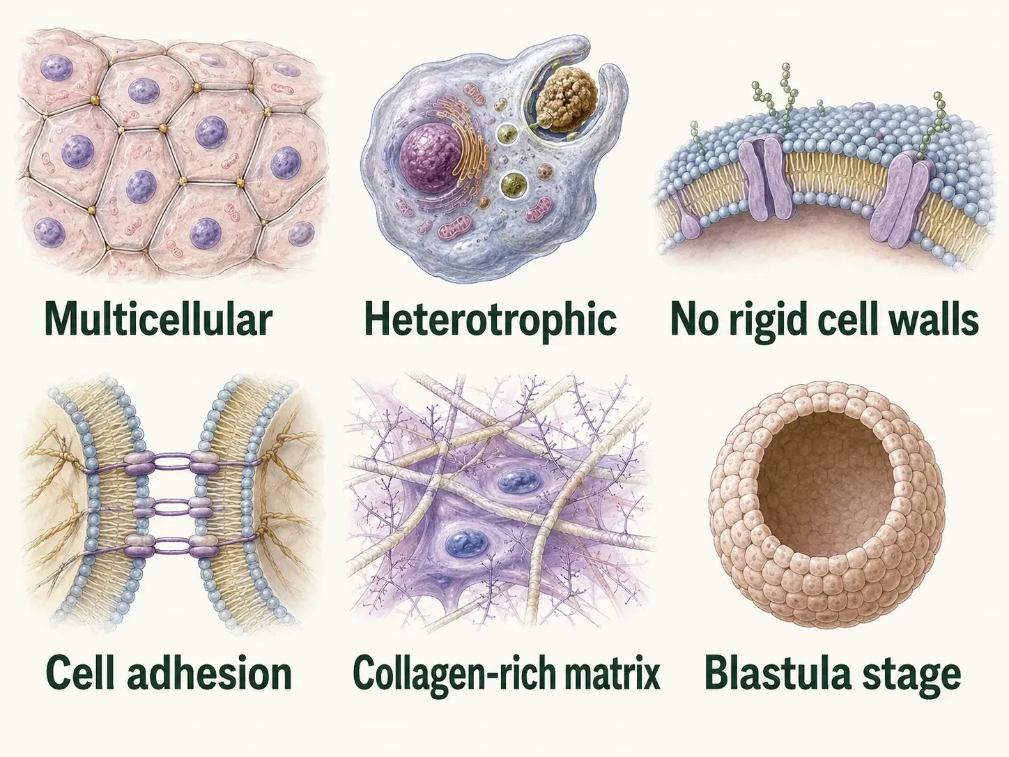
Figure 1. Characteristics of Animals. Animals are multicellular, heterotrophic eukaryotes whose cells lack rigid cell walls and interact through adhesion proteins and a collagen-containing extracellular matrix. Most develop through a blastula stage and possess specialized tissues, although important exceptions occur.
Check Your Understanding Characteristics of Animals
Why is describing animals only as “multicellular heterotrophs” incomplete?
Answer: Multicellularity and heterotrophy are important animal characteristics, but they are not sufficient by themselves. Animals are also eukaryotes whose cells lack rigid cell walls, interact through adhesion systems and an extracellular matrix, and share developmental, molecular, and evolutionary characteristics inherited from a common ancestor.
Why can an organism still be an animal if the adult is sessile or it reproduces asexually?
Answer: Movement and sexual reproduction are common among animals, but neither is universal. Some adult animals remain attached to a surface, and some animals reproduce asexually. Classification depends on shared ancestry and the broader collection of animal characteristics rather than requiring every species to possess every common trait.
Choanoflagellates: The Closest Living Relatives of Animals
Choanoflagellates, sometimes called collared flagellates, are tiny aquatic organisms that are unicellular or exist in simple colonies. They are not animals, but they are the closest living relatives of animals. This makes them especially useful for understanding what the ancestors of animals may have been like.
How do choanoflagellates feed?
A typical choanoflagellate has one long flagellum surrounded by a collar of tiny projections called microvilli. As the flagellum beats, it pulls water toward the cell. Bacteria and other small food particles become trapped along the collar and are then taken into the cell as food. This feeding system is important because it closely resembles the way choanocytes, the feeding cells of sponges, move water and capture food.
Are choanoflagellates multicellular?
Most choanoflagellates live as single cells, but some species can form colonies in which several cells remain attached. In these colonies, cells may communicate and coordinate their behavior, but each cell can still function much more independently than the specialized cells of an animal body. This helps illustrate an important difference between being colonial and being truly multicellular. In animals, different cell types become specialized, depend on one another, and work together as parts of one body; whereas every choanoflagellate cell is identifcal to the next even though they can group together.
What can choanoflagellates tell us about animal origins?
Choanoflagellates and animals share many genes involved in cell adhesion, communication, and gene regulation. These molecular tools existed before the first animals evolved. Early animals later used and expanded this toolkit to help cells stay connected, communicate, and take on specialized roles. Choanoflagellates did not turn into animals, and no living choanoflagellate is the ancestor of modern animals. Instead, choanoflagellates and animals share an ancient common ancestor, and studying both groups helps scientists reconstruct how animal multicellularity evolved.
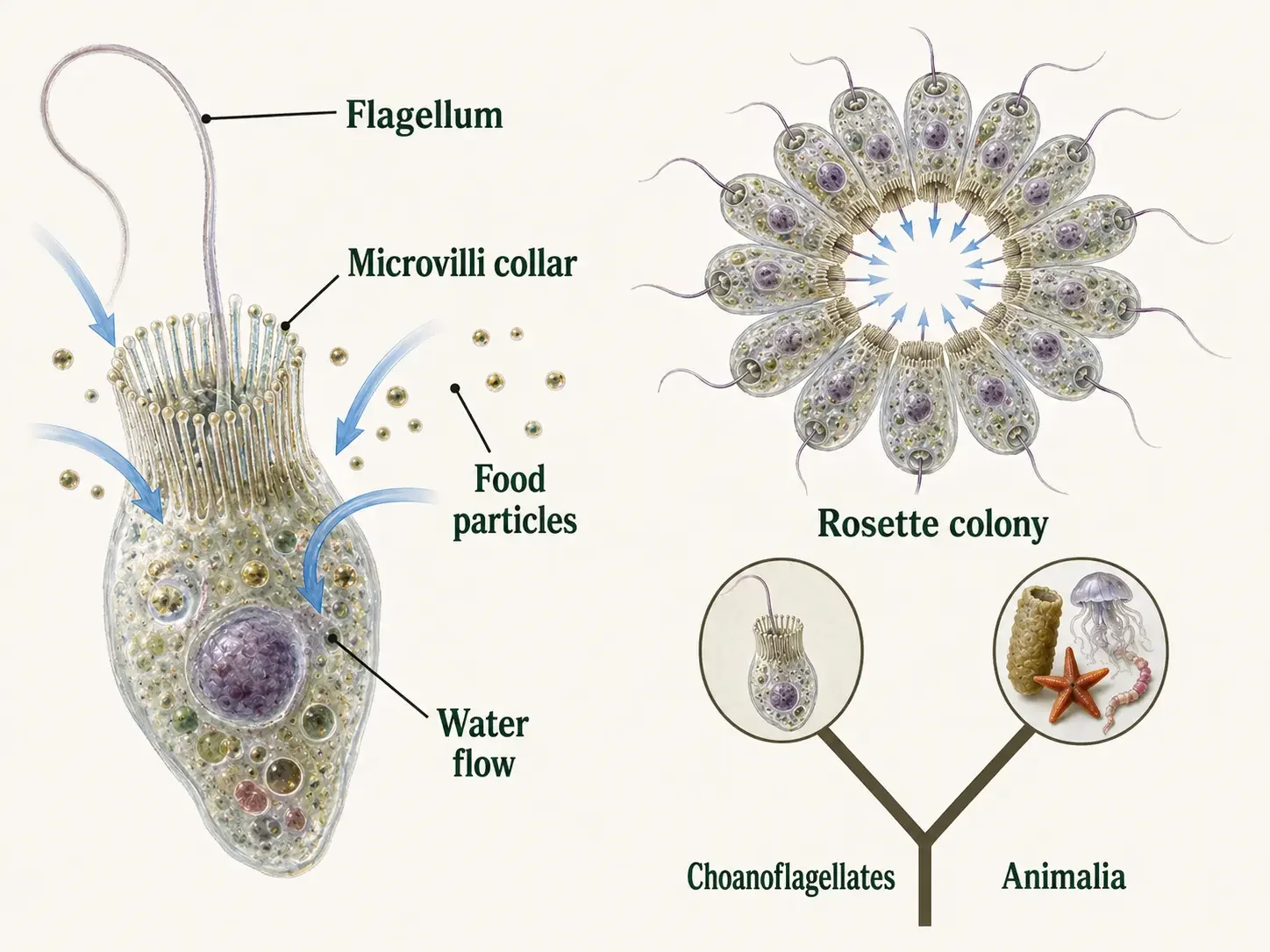
Figure 2. Choanoflagellates and Animal Origins. Choanoflagellates may live as solitary cells or form colonies and feed by drawing water through a collar of microvilli. They are the closest living relatives of animals, not animal ancestors, and share parts of an older molecular toolkit with Animalia.
Animal Origins: How Did Animals Become Multicellular?
The first animals evolved a unicellular species that was also capable of forming simple groups of cells, or colonies. Animals did not evolve from modern choanoflagellates, and living sponges are not unchanged versions of the first animals. Instead, these living groups give us clues about what their ancient ancestors may have been like.
Animal Cells Stick Together
Long before the first animals appeared, their single-celled ancestors already had many of the molecular tools needed for multicellular life. These molecules helped cells stick together, or adhere, allowing neighboring cells to become and remain connected. Adhesion can produce colonial species, like choanoflagellates. Multicellular species require another level of complexity.
Animal Cells Regulate Genes
Ancestors of animals already had molecular systems that could turn genes on and off in response to conditions inside and outside the cell. During the evolution of animal multicellularity, gene regulation allowed genetically identical cells to use different sets of genes and develop different structures and functions. This made cell specialization possible. This is how your skin cell and your bone cell behave differently, eventhough they have the same DNA. When gene regulation worked together with cell adhesion and communication, groups of connected cells could coordinate their activities and function as an integrated multicellular organism.
Animal Cells Communicate
Ancestors of animals already had molecular systems that allowed cells to send and receive signals. During the evolution of animal multicellularity, this communication helped neighboring cells coordinate their behavior and take on different roles. When cell adhesion, communication, and gene regulation worked together, cells could remain connected and function as one interdependent body. This combination of older molecular tools helped make stable animal multicellularity possible.
What did the first animal look like?
We do not know exactly what the first animal looked like, and scientists are still working out the earliest branches of the animal family tree. Strong genomic evidence supports ctenophores (comb jellies) as the sister lineage to all other living animals, although alternative analyses have supported sponges in that position and the question has been historically difficult to resolve. Evolution is a branching tree, not a ladder. Modern choanoflagellates, sponges, ctenophores, and other animals have all been evolving for millions of years, alongside all the other animals. We study them because they preserve different clues about the ancestors they share, not because any living species is a frozen version of the past.
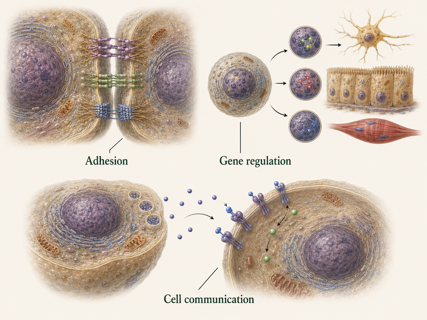
Figure 3. Origin of Animal Multicellularity. Animal multicellularity evolved as existing systems for adhesion, signaling, gene regulation, and extracellular-matrix formation became integrated into stable development. Living choanoflagellates and animals provide comparisons with extinct ancestors, while the earliest branching relationships within Animalia remain an active research problem.
Check Your Understanding Animal Origins and Multicellularity
Why does forming a colony not make a choanoflagellate an animal?
Answer: Cells in a choanoflagellate colony remain much more independent than cells in an animal body. Animal cells become specialized, depend on one another, and work together as parts of one integrated multicellular organism. Choanoflagellates also belong to a separate evolutionary lineage from Animalia.
What do the molecular similarities between choanoflagellates and animals suggest about the origin of animal multicellularity?
Answer: Important molecular tools for cell adhesion, communication, and gene regulation existed before Animalia evolved. During animal evolution, these older tools became integrated and expanded so cells could remain attached, communicate, specialize, and function as one coordinated body.
Why should living choanoflagellates and sponges not be described as unchanged ancestors or evolutionary stages?
Answer: Choanoflagellates and sponges are modern lineages with their own long evolutionary histories. They provide evidence that helps scientists infer characteristics of extinct ancestors, but neither group is a frozen version of the first animal. Evolution forms a branching tree rather than a ladder of progress.
Phylum Porifera: Are they Animals?
Kingdom Animalia → Phylum Porifera
Sponges belong to Phylum Porifera and are true animals, even though they look very different from most other members of Animalia. They are multicellular, their cells work together as a coordinated body, and molecular evidence places them firmly within the animal evolutionary lineage.
What makes sponges multicellular?
This is an important difference between sponges and choanoflagellates. Choanoflagellates are not animals. They are single-celled or colonial relatives of animals. Even when several choanoflagellate cells live together, each cell can function largely on its own. One cell is like all the others, even though they are connected together. In a sponge, however, different cell types are permanently organized to perform different jobs as part of one multicellular animal. This is known as cell differentiation.
Sponges are weird animals!
Sponges have specialized cells that work together to move water, capture food, transport materials, provide support, reproduce, and repair the body. Some sponge cells can change roles when needed, but they still function as parts of one coordinated animal. Sponges possess organized cell layers and some tissue-like features, but they lack the conventional “true tissues” found in most other animals. As a result, they do not form organs such as a heart, brain, or digestive tract, and they have no neurons or true muscle cells.
Characteristics of Sponges
Most sponges are asymmetrical, meaning their bodies cannot be divided into matching halves. Instead of using a mouth and digestive tract, a sponge feeds by moving water through a network of pores and canals. Specialized cells called choanocytes create the water current and capture food particles, while other cells transport nutrients, build skeletal support, and assist with reproduction and repair. Most adult sponges are sessile, meaning they remain attached to a surface. Their larvae are motile and can swim through the water, allowing them to disperse before settling and developing into adults.
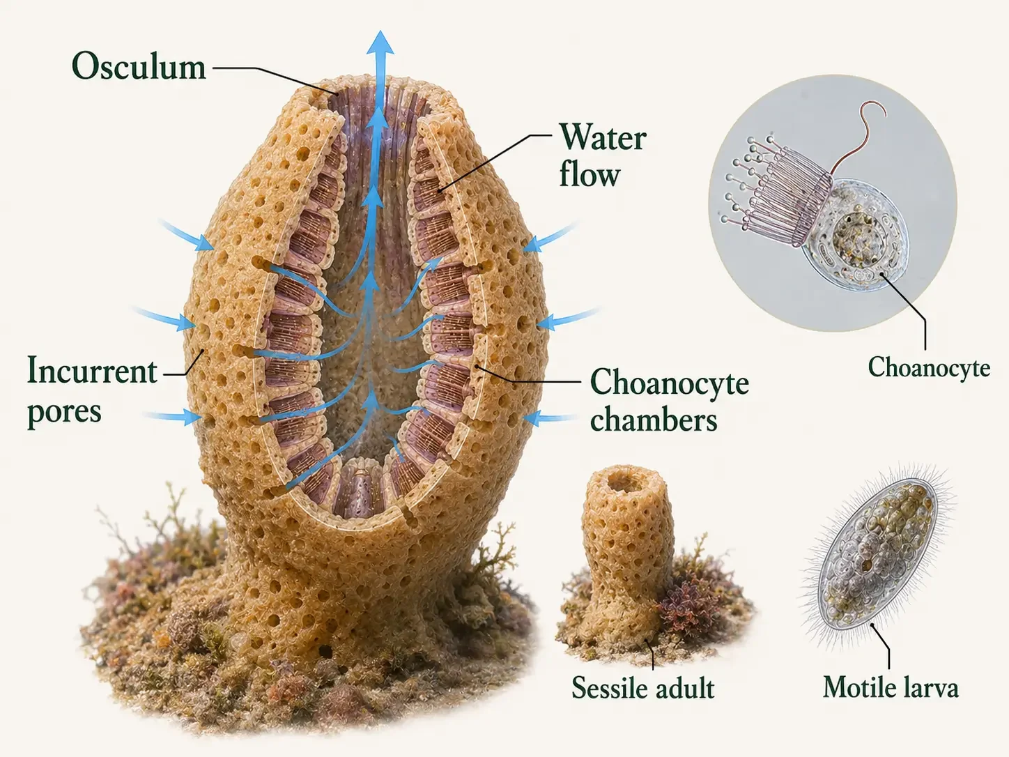
Figure 4. Phylum Porifera. Water enters a sponge through small openings, moves through internal canals and choanocyte chambers, and exits through an osculum. Choanocytes generate the current and capture food, while other specialized cells transport materials, build skeletal support, and contribute to reproduction and repair.
Check Your Understanding Phylum Porifera
Why are sponges classified as animals even though they lack conventional tissues, muscles, nerves, and organs?
Answer: Sponges are multicellular organisms whose specialized cells cooperate as parts of one integrated body. Molecular and phylogenetic evidence also places Porifera firmly within Animalia. The absence of conventional tissues and organs does not make sponges nonanimals or transitional organisms.
How does water move through a sponge, and what role do choanocytes play?
Answer: Water enters through small pores, moves through internal canals and choanocyte chambers, and exits through the osculum. Choanocytes generate the water current and capture suspended food particles. Other specialized cells transport nutrients, provide support, and assist with reproduction and repair.
True Tissues and Functional Layers
Sponges have distinct layers and groups of specialized cells, but these cells are not organized into the stable, integrated tissues found in most other animals. In animals with true tissues, cells are more tightly organized, remain connected, communicate with one another, and work together to perform specific functions.
What is an epithelium?
One common type of animal tissue is the epithelium, a sheet of closely connected cells that forms a continuous surface, generally between tissues. The epithelium helps regulate the movement of substances (i.e water, nutrients, gases, and wastes) between tissues. Epithelia cover the outside of the body and line internal organs, including the digestive tract. You wouldn’t want stomach acid leaking into your stomach cells and beyond. For that, you can thank your epithelium. This kind of organization is one example of what biologists mean by true tissues: cells are not simply grouped together, but are physically connected and work as an integrated unit.
How are sponges different?
Sponge cells on the other hand can change functions as needed. Most other animal cells are fixed. Skin cells are skin cells; bone cells are bone cells. This restriction does not apply to sponges. That is the difference between a sponge tissue and a “true tissue.” While sponges have specialized cells organized into coordinated layers, these layers are not the same as the true tissues found in most other animals. Sponge cells communicate and work together, but they are not organized into the same stable sheets of tightly connected cells seen in tissues such as epithelium. In true tissues, cells are physically joined, maintain a consistent organization, and function together as an integrated unit. Sponges therefore represent a different form of animal body organization rather than a less developed one.
Why do tissues matter?
Tissues allow different groups of cells to perform specialized functions, including protection, movement, digestion, and sensing the environment. Several tissues can also work together to form an organ. This organization supports the many different body plans and ways of life found across animal lineages.
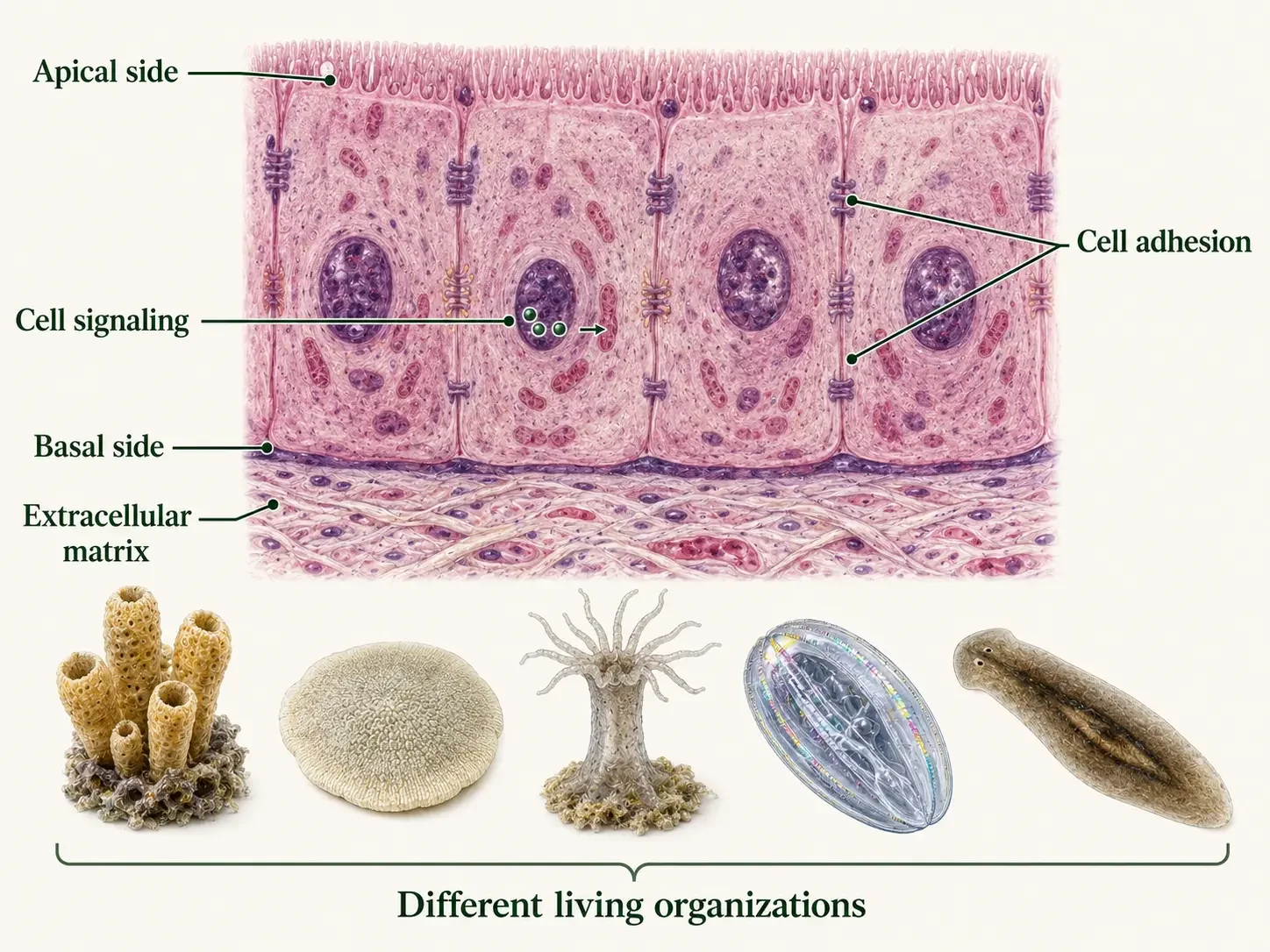
Figure 5. Evolution of Animal Tissues. Tissues depend on cell adhesion, polarity, extracellular matrix, communication, and coordinated cellular functions. Sponges, placozoans, cnidarians, ctenophores, and bilaterians possess different forms of cellular and tissue organization and should be compared as living branches rather than evolutionary stages.
Evolution Note What is Eumetazoa?
Animals with more conventional tissues are often grouped under the term Eumetazoa. Traditionally, this includes cnidarians, ctenophores, and bilaterians, while sponges are placed outside this group.
However, the earliest branches of the animal family tree are still being studied. Because these relationships are not completely settled, Eumetazoa is best treated as a useful biological term rather than as a simple evolutionary step that occurred “after” sponges.
Germ Layers: Building the Body
Early in animal development, the embryo goes through a major reorganization called gastrulation. During gastrulation, cells move into new positions and form layers that will later produce different tissues and body structures. These early layers are called germ layers.
What are the primary germ layers?
Two of the primary germ layers are the ectoderm and endoderm. The ectoderm forms the embryo’s outer layer and later contributes to structures such as the body covering and nervous system. The endoderm forms the inner layer and commonly develops into the lining of the digestive system. Cnidarians are traditionally described as diploblastic because their bodies develop mainly from these two germ layers. In cnidarians, the ectoderm forms the outer epidermis, while the endoderm forms the gastrodermis, which lines a gastrovascular cavity that typically connects to the exterior through a single opening.
What changes in bilaterian animals?
Bilaterians are triploblastic, meaning their embryos develop three primary germ layers: ectoderm, mesoderm, and endoderm. The mesoderm forms between the ectoderm and endoderm. Depending on the animal lineage, mesoderm contributes to muscles, connective tissues, circulatory tissues, reproductive structures, and parts of many internal organs. These tissues allow bilaterians to develop many different internal body arrangements.
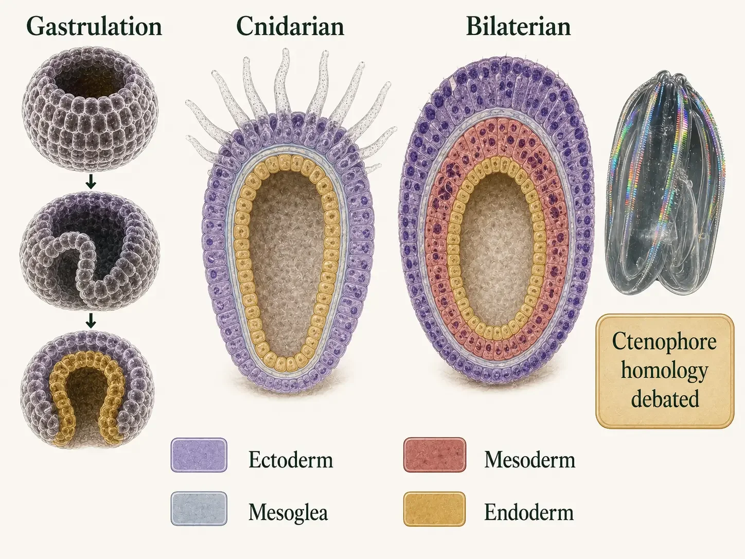
Figure 6. Germ Layers in Animal Development. Gastrulation establishes the primary germ layers. Cnidarians are conventionally described as diploblastic, with ectoderm and endoderm separated by mesoglea, whereas bilaterians are triploblastic and also possess mesoderm. Ctenophore tissues are organized differently, and the homology of their cell layers with the germ layers of other animals remains debated.
Does having three germ layers make an animal “more evolved”?
No. Diploblastic and triploblastic describe patterns of embryonic development, not levels of evolutionary progress.
Evolution is a branching process, so one body plan is not simply a “better” or more advanced version of another.
Body Symmetry: How Is an Animal’s Body Organized?
Body symmetry describes how an animal’s body parts are arranged around its main body axes. Different patterns of symmetry affect how an animal interacts with its environment, moves, and senses what is around it.
What does it mean to be asymmetrical?
Many sponges are asymmetrical, meaning their bodies cannot be divided into matching halves. Their shapes are often irregular because their bodies are built around networks of pores and canals rather than a single, highly organized body axis.
What is radial symmetry?
Many cnidarians, including jellyfish and sea anemones, have radial symmetry. Their body parts are arranged around a central axis, so the animal can interact with the environment from several directions. This body plan works especially well for animals that drift in the water or remain attached in one place. Food, predators, or other environmental signals might approach from almost any direction.
What about ctenophores?
Ctenophores (comb jellies) have a slightly different pattern called biradial symmetry. Their bodies look roughly radial, but only two planes can divide the body into matching halves. This makes their body organization different from the more typical radial symmetry seen in many cnidarians.
What changes in Bilateria?
All other animals are members of Bilateria, which have bilateral symmetry. This means one plane divides the body into a left half and a right half. Bilateral animals also have clear body directions: an anterior end toward the front, a posterior end toward the back, a dorsal side toward the back or upper surface, and a ventral side toward the belly or lower surface. This organization is often associated with moving through the environment in a particular direction.
Why does symmetry matter?
Symmetry is more than just the shape of an animal. It reflects how the body is organized to move, feed, sense the environment, and respond to threats. Radial and biradial animals are well suited for interacting with the world from many directions. Bilateral symmetry, in contrast, is often linked with directional movement and a more clearly defined front end.
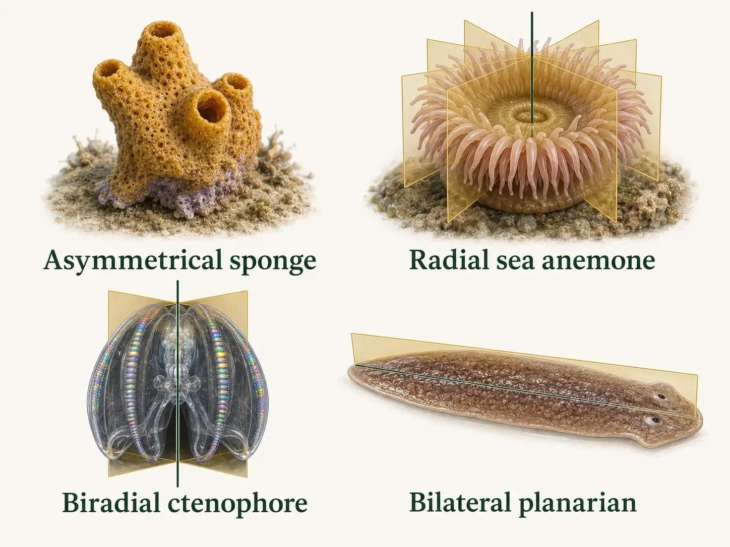
Figure 7. Body Symmetry in Animals. Many sponges are asymmetrical, many cnidarians exhibit radial symmetry, ctenophores exhibit biradial symmetry, and bilaterians ancestrally possess bilateral symmetry. These are different patterns of body organization—not stages in a progression—and they may be modified within individual animal lineages.
Check Your Understanding Germ Layers and Body Symmetry
Diploblastic Animals: What Does “Diploblastic” Really Mean?
Animal Body Plans → Germ Layers
Some animals are described as diploblastic, meaning their embryos develop mainly from two primary germ layers: the ectoderm and endoderm. These layers later form different tissues and structures in the adult body.
Which animals are diploblastic?
Cnidarians, including jellyfish, corals, and sea anemones, are conventionally described as diploblastic. Their bodies develop mainly from an outer ectoderm and an inner endoderm. Ctenophores (comb jellies) have also traditionally been called diploblastic. However, their development includes mesoderm-like cell lineages, and scientists are still studying how these cells compare with the true mesoderm found in bilaterian animals.
Why is this distinction important?
It is easy to imagine animal evolution as a simple sequence: sponges first, then diploblastic animals, then triploblastic animals. But evolution does not work like a ladder. Terms such as diploblastic and triploblastic are useful for describing how animal bodies develop. They should not be treated as steps showing that one group is “more evolved” than another.
Does diploblastic describe a branch of the animal family tree?
No. Diploblastic describes a pattern of development, not a formal evolutionary group.
Cnidarians and ctenophores belong to two separate animal phyla: Cnidaria and Ctenophora.
Older classification systems sometimes grouped these animals together as Radiata, but modern evolutionary evidence does not support Radiata as a single natural clade.
Phylum Cnidaria: The Animals with Stinging Cells
Kingdom Animalia → Phylum Cnidaria
Cnidarians include jellyfish, corals, hydroids, and sea anemones. Their defining feature is a special cell called a cnidocyte, which helps them capture prey and defend themselves. Inside each cnidocyte is a tiny capsule. The best-known type is a nematocyst, which can rapidly fire a thin thread that may pierce, tangle, or stick to another organism. Many nematocysts also release venom.
How is a cnidarian body built?
Cnidarians have two main tissue layers. The outer epidermis (ectoderm → epidermis) covers the body, while the inner gastrodermis lines the digestive system (endoderm → gastrodermis). Food is digested inside a gastrovascular cavity, which usually has one opening that acts as the mouth and the anus simultaneously.
Do cnidarians have a brain?
Cnidarians do not have a centralized brain like many bilaterian animals. Instead, most have a network of neurons called a nerve net, which receives and processes sensory information and coordinates behaviors such as swimming, feeding, and escaping danger. Some cnidarians also have clusters of neurons and sensory structures that detect light, chemicals, touch, gravity, or body position. Their nervous systems are organized differently from ours, but they do more than simply sense and react.
What are polyps and medusae?
Cnidarians commonly have two body forms. A polyp is usually attached to a surface, while a medusa is usually free-swimming. Jellyfish are medusae, while corals and sea anemones are polyps. Some cnidarians switch between these forms during their life cycle, but not all do.
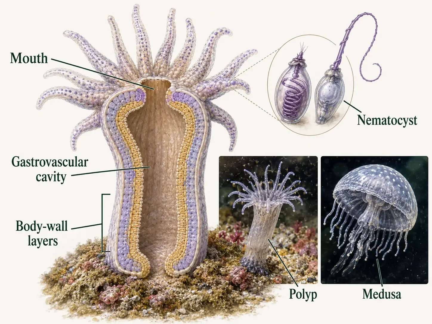
Figure 8. Phylum Cnidaria. Cnidarians possess an epidermis and gastrodermis separated by mesoglea, and their gastrovascular cavity typically opens through a single mouth. Their defining cnidocytes contain cnidae; the illustrated nematocyst is a capsule that rapidly everts a thread. Polyp and medusa forms vary across jellyfish, sea anemones, corals, and hydroids.
How do corals build reefs?
Many corals produce hard calcium carbonate skeletons. As generations of corals grow, die, and build on older skeletons, this material gradually accumulates.
Over long periods of time, these accumulated skeletons help form the massive structures we know as coral reefs.
Phylum Ctenophora: The Comb Jellies
Kingdom Animalia → Phylum Ctenophora
Ctenophores, or comb jellies, are soft, jelly-like marine animals that belong to a different lineage from cnidarians. Most have biradial symmetry, meaning their bodies can be divided into matching halves along two planes.
How do comb jellies move?
Ctenophores swim using eight rows of structures called comb plates. Each comb plate is made of many fused cilia, tiny hair-like structures that beat together in waves. These moving rows often reflect light, creating the shimmering rainbow effect seen in many comb jellies.
How do ctenophores catch food?
Many ctenophores use long tentacles covered with colloblasts, special sticky cells that help trap small prey. Unlike cnidarians, ctenophores do not use stinging cnidocytes. Some species do not have tentacles at all and capture prey directly with their mouths.
How does digestion work?
Food enters through the mouth, passes through a short throat-like region, and moves into a branching digestive canal system. Nutrients are distributed through these canals, and waste leaves through small anal pores near the opposite end of the body.
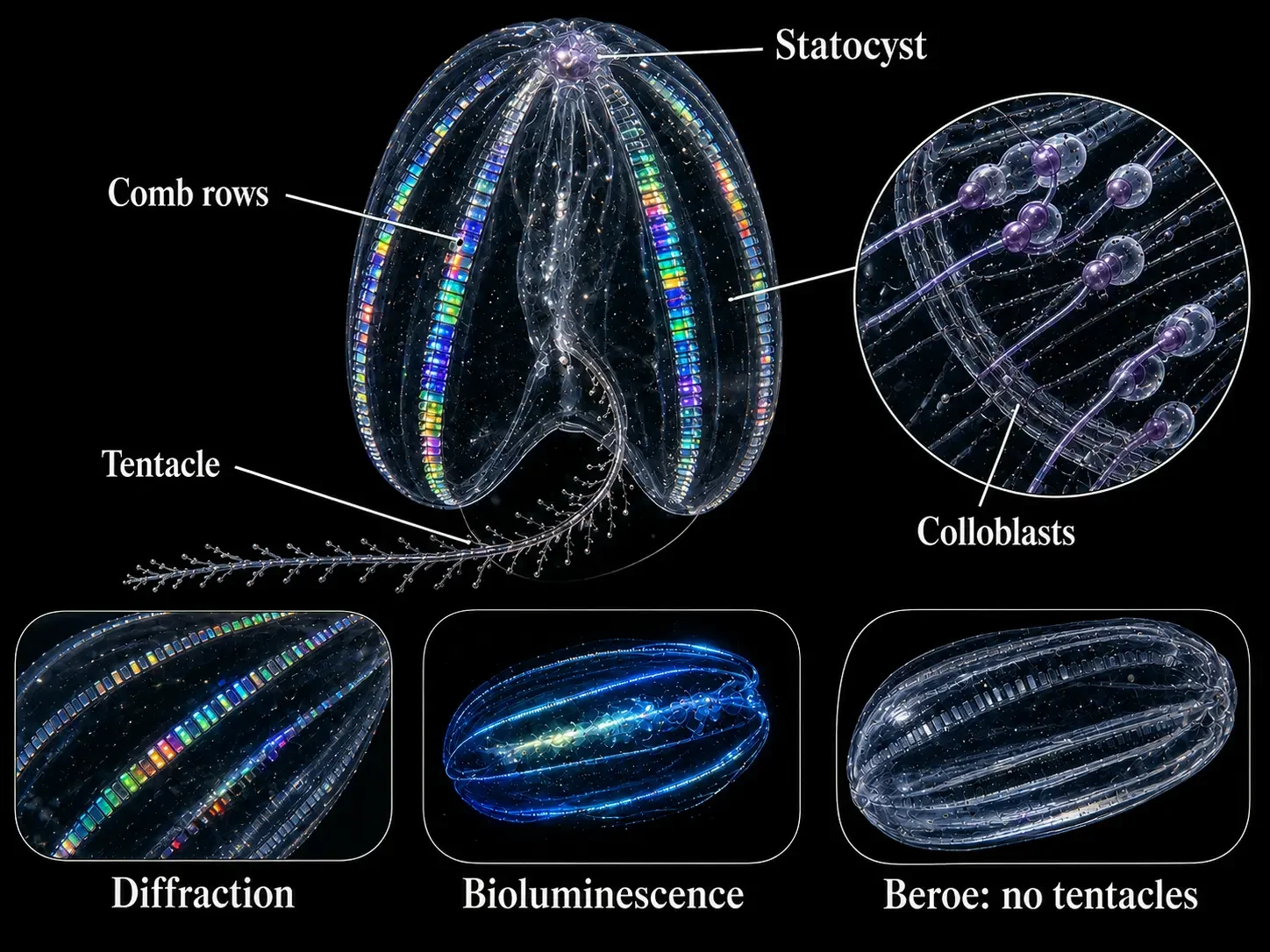
Figure 9. Phylum Ctenophora. Ctenophores move with eight rows of fused ciliary plates called combs and orient using an aboral statocyst. Many capture prey with sticky colloblasts on tentacles, although genera such as Beroe lack tentacles. Rainbow colors along the comb rows result from diffraction and are distinct from bioluminescence.
Check Your Understanding Cnidaria and Ctenophora
Florida Man Pro Tip Where to go see bioluminescence in Florida?
if you are near Cape Canaveral, skip the crowded beach after sunset and head toward the quiet water on the other side of the barrier island. The Indian River Lagoon system can contain thousands of tiny, gelatinous animals that flash electric blue-green light when the water around them is disturbed.
🌌 When should you go?
If your goal is specifically to see bioluminescent ctenophores, plan your trip for the cooler part of the year.
The exact abundance changes from year to year, so think of this as a biological season rather than an appointment. Cooler months favor the comb-jelly experience, while Florida's famous summer bioluminescence is usually dominated by microscopic dinoflagellates.
🌑 Florida Man's secret weapon: check the Moon
Go on a dark night. Ideally, plan your paddle within several nights of the new moon. Less moonlight means your eyes can detect much fainter flashes in the water.
Give your eyes time to dark-adapt. Avoid shining bright white flashlights across the water, and turn your phone screen brightness down before launching.
Cool winter night + new moon + calm water + little artificial light = your best chance at a spectacular ctenophore show.
📍 Where should you go?
For bioluminescent ctenophores, the real Florida hotspot is not the open Atlantic Ocean. It is the protected lagoon system behind the Space Coast: the Indian River Lagoon, Mosquito Lagoon, and Banana River.
-
🥇 Haulover Canal — Merritt Island National Wildlife Refuge
Best overall choice. North of Kennedy Space Center near Titusville, Haulover Canal connects Mosquito Lagoon with the northern Indian River Lagoon. The surrounding refuge has very little artificial lighting, making this one of the strongest combinations of dark sky + protected water + ctenophore habitat.
Florida Man rating: 🐊🐊🐊🐊🐊
-
🥈 Beacon 42 — Merritt Island National Wildlife Refuge
Another excellent Titusville-area launch within the refuge. Beacon 42 gives paddlers access to dark sections of the lagoon with very little surrounding development. It is particularly impressive when the sky itself is dark enough to see large numbers of stars.
Florida Man rating: 🐊🐊🐊🐊🐊
-
🥉 Banana River — Merritt Island / Cocoa Beach
The Banana River is an excellent option if you are staying near Cocoa Beach, Cape Canaveral, or Port Canaveral. Protected stretches of the river can support both winter comb jellies and warmer-season dinoflagellate bioluminescence.
Florida Man rating: 🐊🐊🐊🐊
-
Kiwanis Island Park — Merritt Island
A convenient Banana River access point near Cocoa Beach. This is a good choice when you want the bioluminescence experience without driving as far north into the wildlife refuge.
Florida Man rating: 🐊🐊🐊🐊
-
Kelly Park East — Merritt Island
Another Banana River access point close to Port Canaveral and Cape Canaveral. It is convenient and commonly used for bioluminescence excursions, although conditions vary seasonally.
Florida Man rating: 🐊🐊🐊½
🚀 The Cape Canaveral strategy
If you are visiting Kennedy Space Center, you are already in the right neighborhood. Spend the day exploring the Space Coast and schedule a nighttime paddle near Haulover Canal or Beacon 42.
You may literally spend the afternoon looking at spacecraft and the evening watching an animal lineage more than half a billion years old produce its own light.
🔦 How do you actually see them glow?
Look down beside the kayak and gently move your paddle through the water. When disturbed, bioluminescent ctenophores may flash blue-green. A passing fish can occasionally produce a glowing streak as it disturbs organisms in the water.
In daylight, you may also notice shimmering rainbow colors moving along a ctenophore's eight rows of comb plates. That rainbow effect is not bioluminescence. It results from light interacting with the rows of beating cilia. The blue-green flash visible at night is the animal's actual bioluminescence.
Want glowing water everywhere? Come in summer for dinoflagellates.
Want to see an actual animal flash with bioluminescence? Come in winter and hunt for ctenophores.
For the best shot: November–March + new moon + Merritt Island National Wildlife Refuge + Haulover Canal or Beacon 42.
Bilateria: Animals with a Left and Right Side
Kingdom Animalia → Bilateria
Bilateria is the large animal group whose members are built around a clear front-to-back body axis. Most bilaterians also have bilateral symmetry, meaning one plane can divide the body into left and right halves.
Triploblasty: What makes bilaterians different?
Bilaterians are triploblastic, meaning their embryos develop three primary germ layers: ectoderm, mesoderm, and endoderm. These layers later form different tissues and organs, giving bilaterians more ways to build specialized body structures.
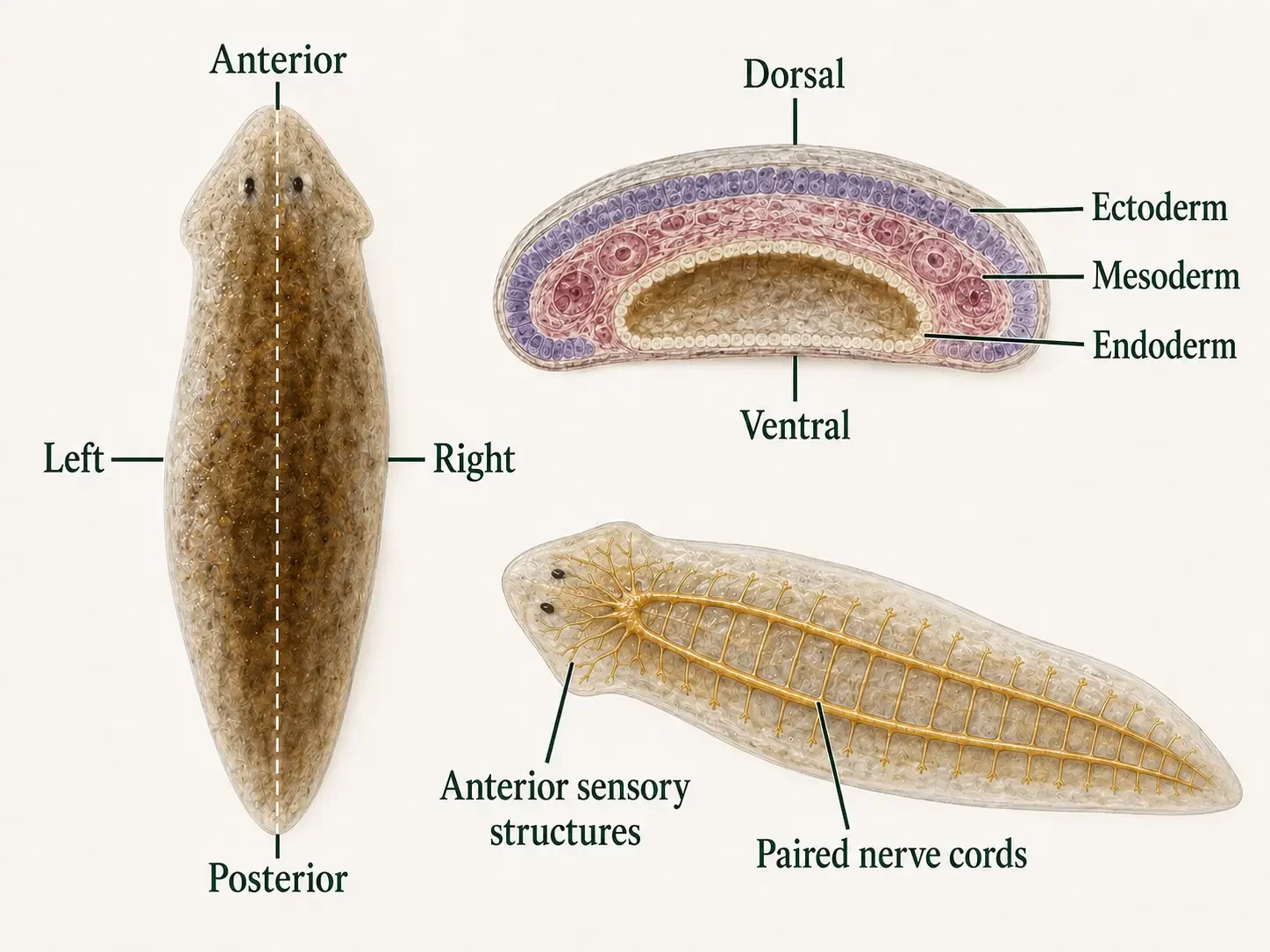
Figure 10. Bilateria. Bilaterians ancestrally possess left and right sides, distinct anterior and posterior ends, and dorsal and ventral surfaces. Their embryos form ectoderm, mesoderm, and endoderm, and many lineages concentrate sensory structures and nervous tissue toward the anterior through cephalization. These ancestral patterns can be modified in descendant lineages.
Exception to the Rule: Not all bilaterians are bilateral
Most bilaterians retain the basic left-right body plan, but some groups have modified it during evolution.
Echinoderms, such as sea stars, are a good example: their larvae are bilaterally symmetrical, while the adults develop a more radial body shape.
Mesoderm: The Third Germ Layer
Kingdom Animalia → Bilateria
Bilaterian embryos develop three primary germ layers: ectoderm, mesoderm, and endoderm. The ectoderm forms the outer covering and much of the nervous system, while the endoderm forms the lining of the digestive tract and some associated organs. The mesoderm develops between them and is what makes bilaterians triploblastic.
What does mesoderm form?
The mesoderm contributes to many structures inside the body, including muscles, connective tissues, reproductive structures, circulatory tissues, and many internal organs. The exact structures it forms differ among animal groups, but mesoderm gives the body more ways to organize tissues between the outer body wall and the digestive tract.
Why was mesoderm important?
With a third germ layer, animals could develop new arrangements of muscles, organs, and body cavities. This gave bilaterian animals more flexibility in how their bodies were built and how different internal structures could work together.
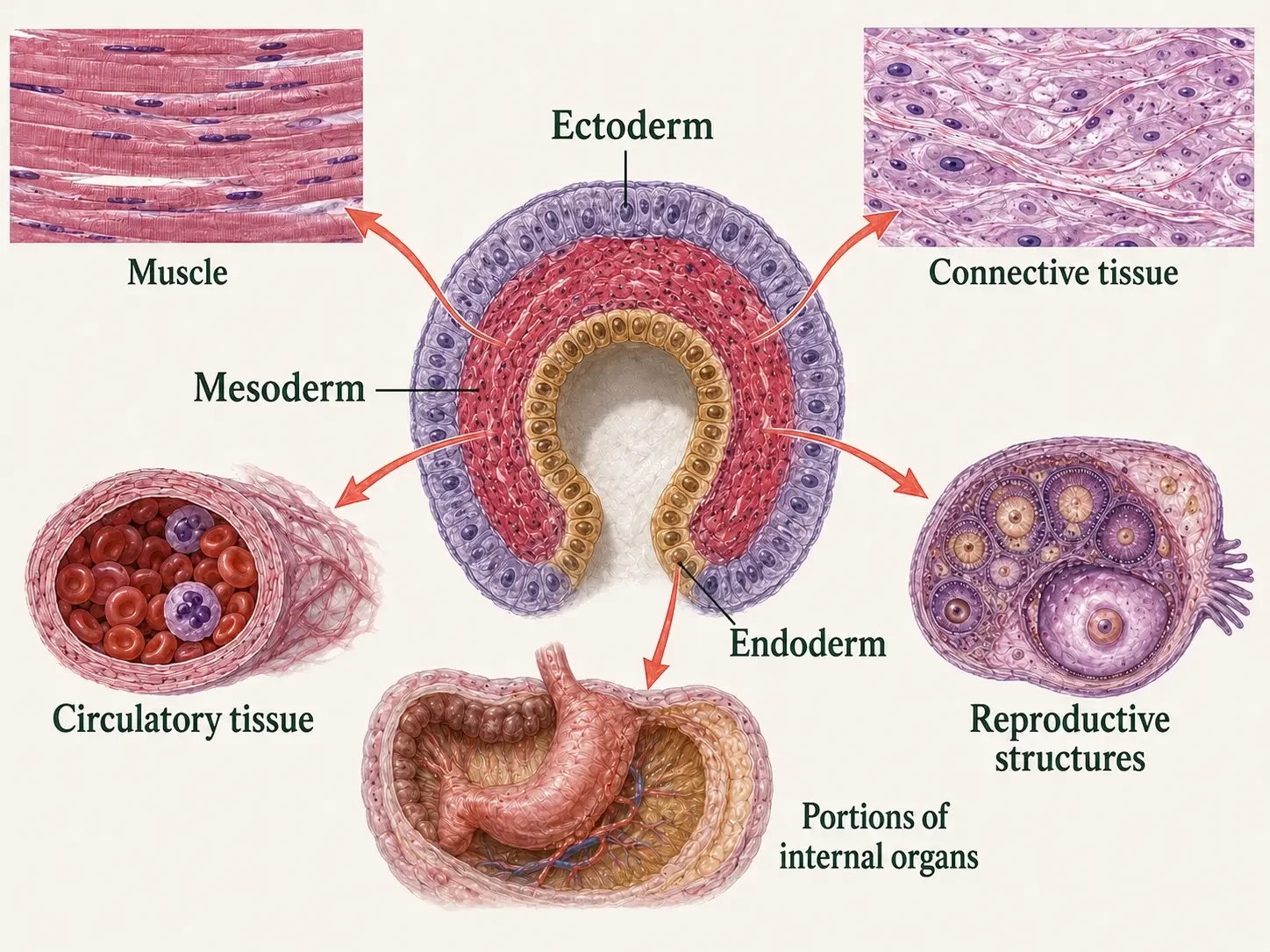
Figure 11. Origin of Mesoderm. Mesoderm forms between ectoderm and endoderm during bilaterian development. Depending on the lineage, mesoderm contributes to muscle, connective and circulatory tissues, reproductive structures, and portions of internal organs; the specific derivatives vary among animals.
Directional Movement Can Favor Cephalization
Kingdom Animalia → Bilateria
Bilaterians have a clear front and back, as well as left and right sides. In animals that usually move forward, the front end encounters new surroundings first. Over time, natural selection can favor placing important sensory structures near the front, where they can quickly detect food, predators, obstacles, and potential mates.
Get a head: Why cephalization?
Cephalization is the concentration of sensory structures, feeding structures, and nervous tissue near the front end of the body. This organization is especially useful in bilaterians that usually move in one main direction because the front end encounters new surroundings first. Grouping sensory structures and nervous tissue there can help an animal detect food or danger, process that information, and respond quickly. Greater centralization can reduce the delay between sensing a change in the environment and reacting to it.
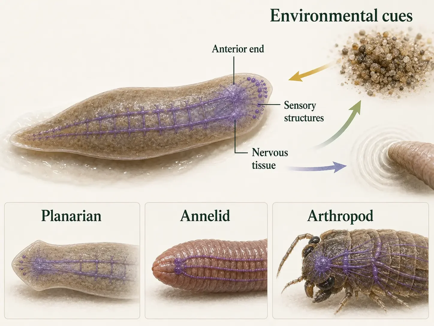
Figure 12. Bilateral Symmetry and the Head Region. An animal moving in a consistent direction encounters environmental cues at its anterior end first. Cephalization concentrates sensory structures and nervous tissue in that region, but its form and degree vary widely among bilaterian lineages.
Check Your Understanding Bilateral Symmetry and the Head Region
Why can an adult sea star have radial organization and still belong to Bilateria?
Answer: Bilateria is an evolutionary lineage, not a requirement that every adult animal retain bilateral symmetry. Sea star larvae possess bilateral symmetry, but adults develop a derived five-part radial organization. This demonstrates that ancestral symmetry patterns can be modified within an animal lineage.
Acoelomorpha: Bilaterians Without a Body Cavity
Kingdom Animalia → Bilateria → Acoelomorpha
Acoelomorpha are small, soft-bodied bilaterian animals that live mostly in marine environments. Like other bilaterians, they have bilateral organization and tissues derived from mesoderm, but their bodies do not contain a fluid-filled body cavity called a coelom.
How is an acoelomorph body organized?
Acoelomorphs have compact bodies, with tissues filling most of the space between the body wall and the digestive region. Their digestive systems vary among groups. Many acoelomorphs do not have a permanent hollow gut; instead, food is digested within a central mass of cells. Other members have a simple digestive cavity, but they generally lack a separate anus, so undigested material leaves through the same opening used to take in food.
Where do acoelomorphs fit on the animal tree?
Scientists are still figuring out exactly where these animals fit on the bilaterian family tree. Acoelomorphs are small, worm-like animals that are often placed in a group called Xenacoelomorpha. Some studies suggest that this group branched off early in bilaterian evolution, while others place it closer to echinoderms and their relatives. Where the group belongs matters because it can change how scientists explain the early evolution of bilaterian bodies. These animals are living species today, not ancient ancestors or evolutionary halfway stages.
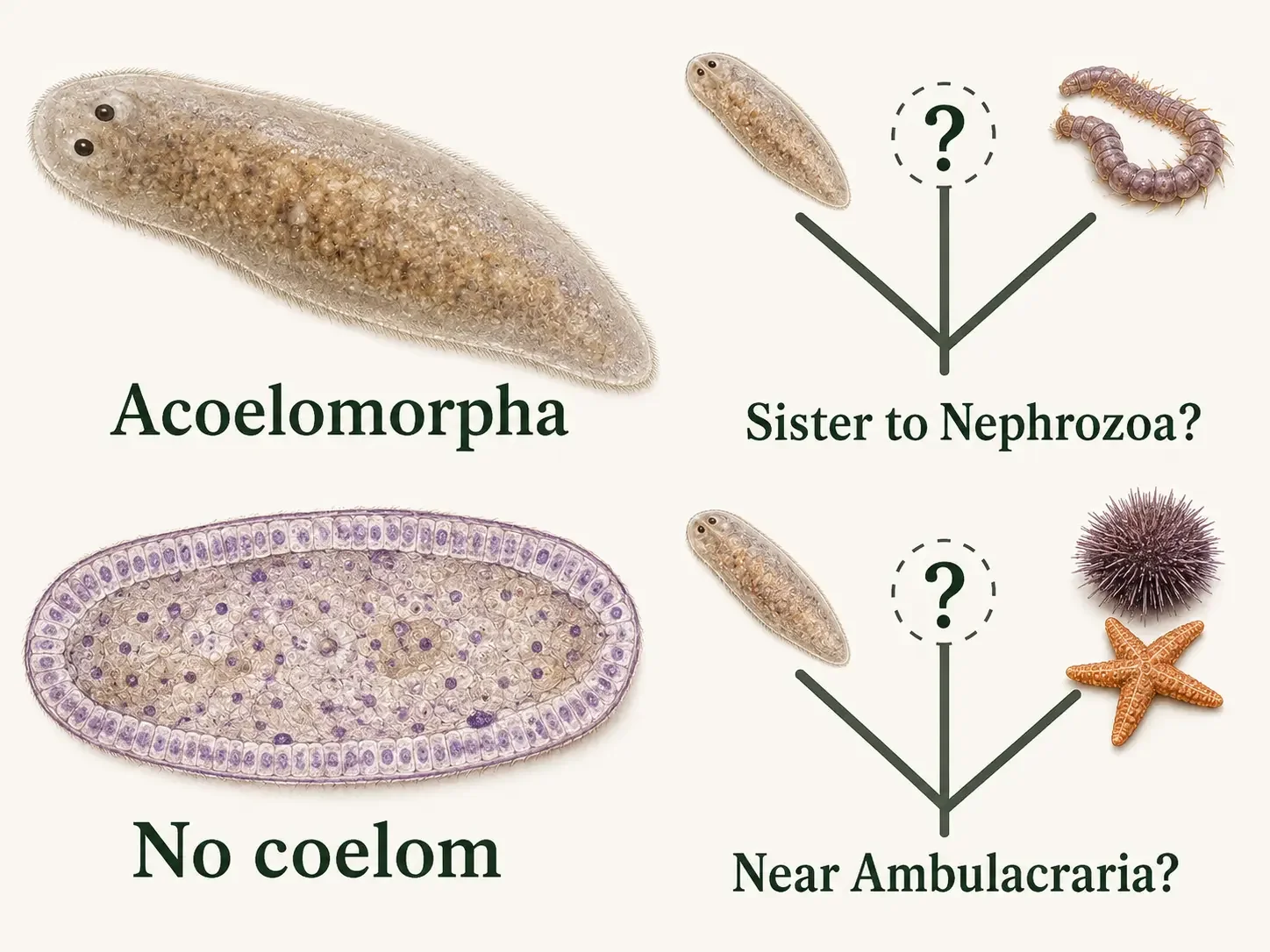
Figure 13. Acoelomorpha. Acoelomorphs are small bilaterian animals with compact bodies, mesoderm-derived tissues, and no coelom. Their simplicity does not make them primitive or direct ancestors of other animals. The phylogenetic placement of Xenacoelomorpha remains debated, with competing hypotheses placing it outside Nephrozoa or near Ambulacraria.
The Coelom: Moving More Efficiently
A coelom is a fluid-filled space between the digestive tract and the outer body wall. It is different from simply having an empty space inside the body. A true coelom is completely lined by mesoderm, the middle germ layer that also gives rise to structures such as muscles and connective tissues. This mesodermal lining helps support and organize the organs that sit within the cavity.
Why is a coelom useful?
A coelom gives internal organs more room to grow, move, and function without being packed tightly against the body wall. This can allow the digestive system and other organs to move somewhat independently from the rest of the body. The fluid inside can also help support the body, distribute forces, cushion organs, and move materials over short distances. In some animals, the coelomic fluid even works with muscles as part of a hydrostatic skeleton, helping the animal maintain its shape and move.
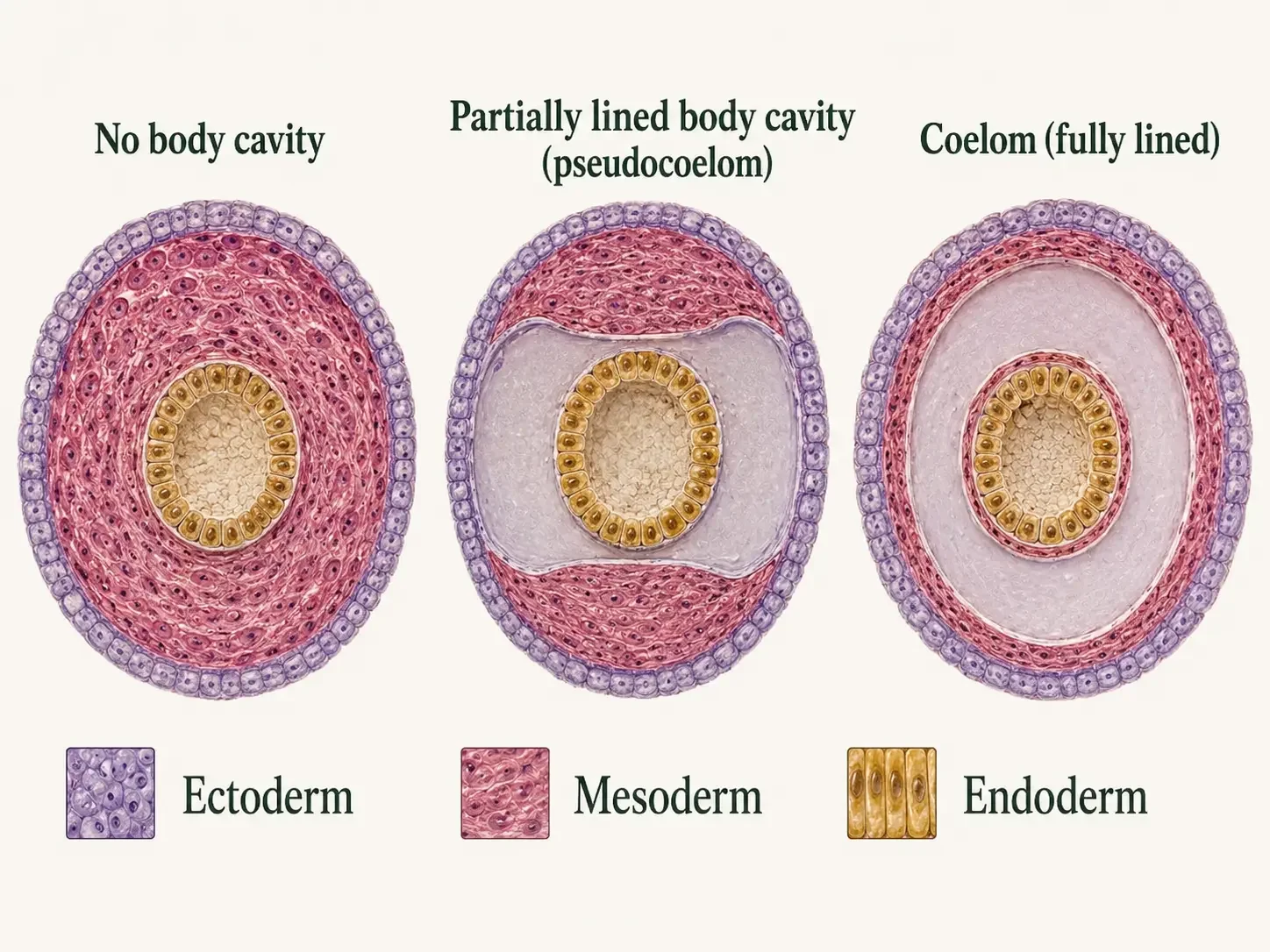
Figure 14. Evolution of the Coelom. A coelom is a fluid-filled body cavity fully lined by mesoderm. Animal lineages also possess compact body plans without a body cavity or cavities that are not fully lined by mesoderm. These arrangements can create space, cushion organs, and contribute to support, but they are alternative body plans rather than stages of advancement.
Are acoelomorphs an evolutionary “step” toward animals with coeloms?
No. Acoelomorphs are not a halfway stage between animals without mesoderm and animals with body cavities.
They are modern animals with their own long evolutionary history. Their relatively simple body organization represents adaptations that evolved along their own branch of the animal tree.
Hydrostatic Skeletons: Coeloms Allow Efficient Movement
In many soft-bodied animals, fluid inside the body can act as part of a hydrostatic skeleton. Because fluid is hard to compress, muscles can push against it to change the animal’s shape and produce movement without needing bones.
How does a hydrostatic skeleton work?
When muscles contract around a fluid-filled space, the fluid pushes back and transfers that force through the body. This allows one part of the body to become longer, shorter, wider, or narrower while the overall volume stays nearly the same.
How does an earthworm use one?
An earthworm is a familiar example. Circular muscles make a body segment longer and thinner, while longitudinal muscles make it shorter and wider. Small bristles called setae help anchor parts of the worm as waves of muscle contraction move along the body. Internal partitions between segments help keep fluid pressure more localized. This gives the worm better control over which parts of the body change shape at a given time and makes movement more efficient.
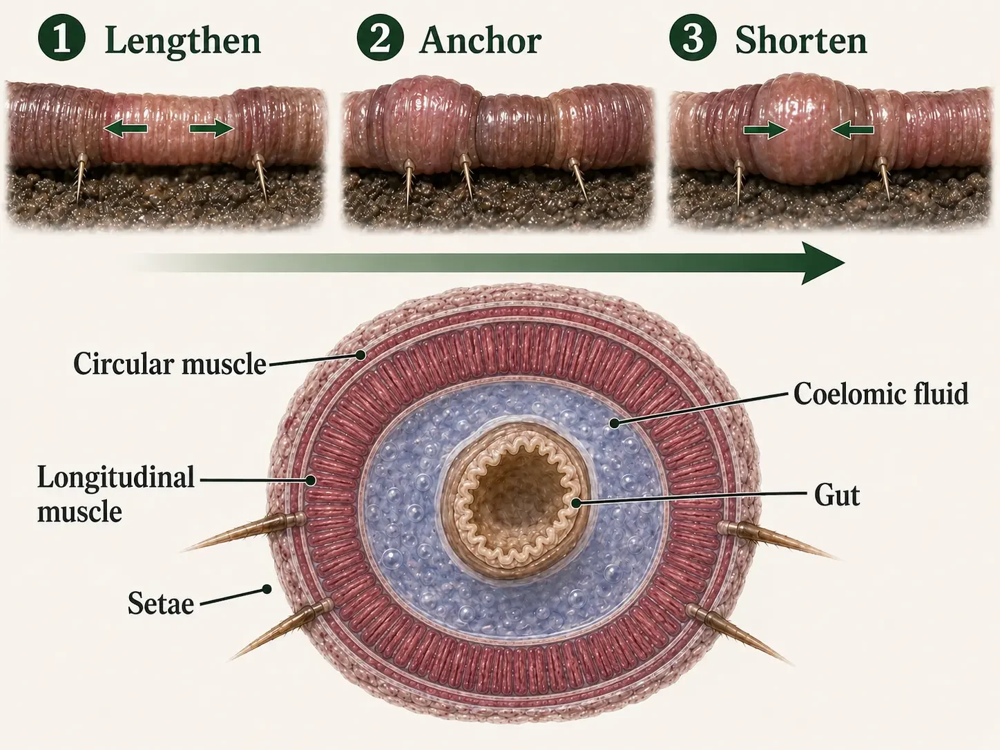
Figure 15. Coelom and Hydrostatic Movement. In an earthworm, circular muscles lengthen and narrow segments while longitudinal muscles shorten and widen them. Setae anchor selected segments, and waves of alternating contraction act against incompressible coelomic fluid to move the animal forward.
Check Your Understanding The Coelom and Hydrostatic Movement
Protostomia and the Traditional Deuterostome Grouping
Kingdom Animalia → Bilateria → Nephrozoa
Most familiar bilaterian animal groups are commonly placed within Nephrozoa. One strongly supported branch, Protostomia, contains two major lineages: Spiralia and Ecdysozoa. Spiralia includes flatworms, rotifers, annelids, mollusks, and related animals. Ecdysozoa includes animals that molt an external covering, such as nematodes and arthropods. Echinoderms, hemichordates, and chordates have traditionally been grouped as Deuterostomia. However, some recent phylogenomic studies question whether Deuterostomia forms a single natural evolutionary branch. These animals are introduced here and examined more closely in the next two chapters.
How were these animals originally compared?
Protostomes and the traditional deuterostome groups were historically compared using patterns observed during embryonic development. Biologists examined how early embryonic cells divide, what happens to the blastopore, and how the coelom forms. We will dive into these concepts next. These patterns remain useful for learning, but they are general tendencies rather than rules. No single developmental pattern occurs in every member of either group.
How do scientists classify these animals today?
Modern classification combines evidence from anatomy, embryonic development, DNA, and entire genomes. This evidence strongly supports Protostomia as a major evolutionary lineage and has clarified many relationships within Bilateria. However, the relationships among chordates, echinoderms, and hemichordates remain under investigation, and some studies do not recover Deuterostomia as one natural evolutionary group. The traditional comparison remains useful as long as its developmental patterns are treated as tendencies and its phylogeny is presented as an active area of research.
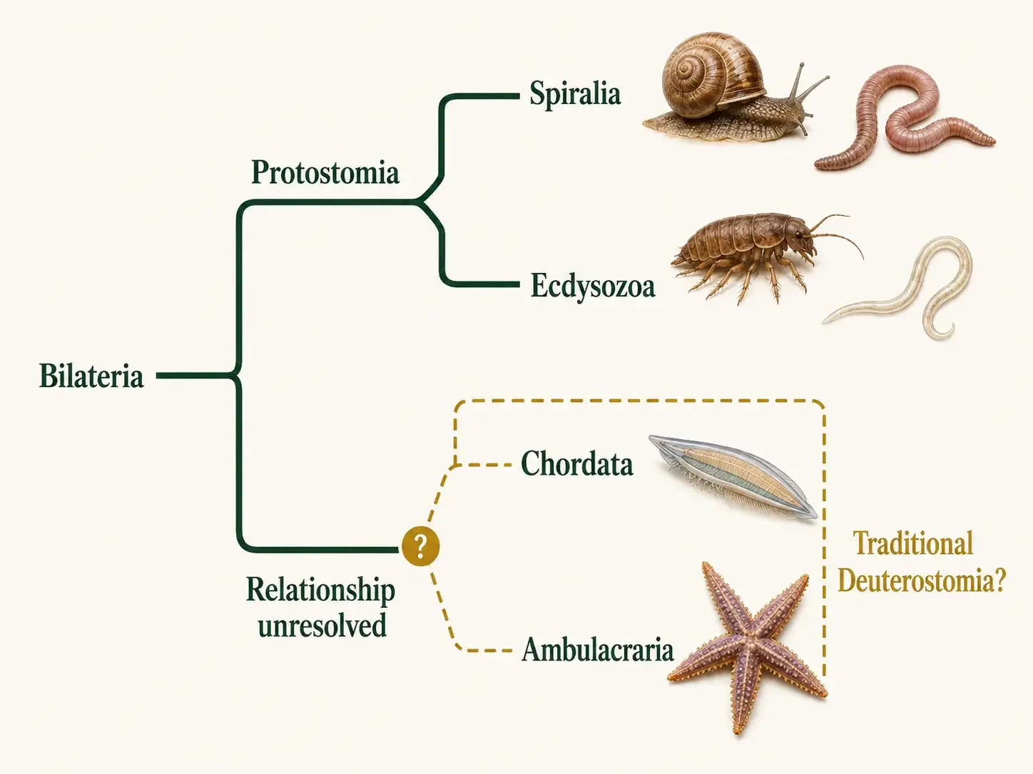
Figure 16. Protostomia and the Traditional Deuterostome Grouping. Protostomia is a strongly supported clade that includes Spiralia and Ecdysozoa. Chordata and Ambulacraria have traditionally been grouped as Deuterostomia, but some phylogenomic analyses question whether that grouping is monophyletic. The unresolved branch geometry presents this as a teaching framework rather than a settled claim.
Blastopore Fate: A Useful Pattern, Not a Rule
During gastrulation, cells move inward and form an opening in the embryo called the blastopore. Imagine pushing a finger into a balloon. That is akin to a blastopore. What happens to this opening became one of the classic ways biologists compared protostomes and deuterostomes.
What happens to the blastopore?
In the traditional pattern, the blastopore becomes the mouth in many protostomes. In many animals traditionally grouped as deuterostomes, it becomes the anus. This difference is where the names protostome and deuterostome originally came from. In Greek, protostome means “first mouth” (proto = first, stoma = mouth), while deuterostome means “second mouth” (deutero = second, stoma = mouth). The terms were originally based on what scientists thought happened to the first opening formed during early embryonic development.
Do all protostomes and deuterostomes follow this pattern?
No. Animal development is more complicated. Depending on the species, the blastopore may contribute to one digestive opening, contribute to both openings, or close while new openings form elsewhere. Blastopore fate remains useful for comparing broad developmental patterns, but it is a general tendency rather than a rule for determining whether an animal belongs to Protostomia or to a lineage traditionally grouped as Deuterostomia.
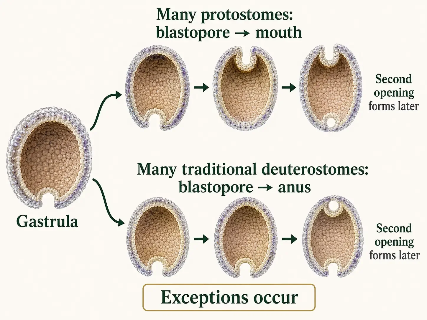
Figure 17. Blastopore Fate. In the classical pattern, the blastopore contributes to the mouth in many protostomes and to the anus in many lineages traditionally called deuterostomes; a second opening forms the other end of the digestive tract. Numerous exceptions make blastopore fate a developmental tendency, not a universal diagnostic trait.
Early Cleavage: Different Patterns in Early Embryos
Early animal embryos grow by going through a series of rapid cell divisions called cleavage. These divisions split the embryo into smaller cells without greatly increasing its overall size.
What is spiral cleavage?
In spiral cleavage, new cells form at slight angles to the cells below them, creating a twisting pattern. This is especially common in many spiralian protostomes, such as annelids and mollusks. In these animals, individual cells also begin taking on specific developmental roles fairly early. Ecdysozoans, including arthropods and nematodes, develop in different ways, so the simple spiral-versus-radial comparison should not be applied to every protostome.
What is radial cleavage?
In radial cleavage, early cells tend to line up more directly above one another in rows or tiers. This pattern is common in many animals traditionally grouped as deuterostomes, and their early cells often keep the ability to form a wider range of tissues for a longer time.
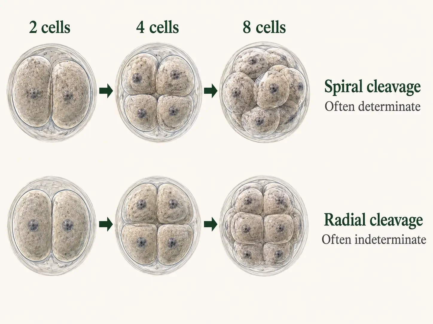
Figure 18. Early Embryonic Development. Spiral, often determinate cleavage is common in many spiralians but is not universal among protostomes. Radial, often indeterminate cleavage is common in many traditional deuterostome lineages. Cleavage patterns and developmental potential vary among lineages, and exceptions occur.
Coelom Formation: Different Ways to Make a Body Cavity
A coelom is a fluid-filled body cavity lined by mesoderm. During development, animals can form this cavity in different ways. Two well-known patterns are schizocoely and enterocoely.
What is schizocoely?
In schizocoely, the mesoderm first forms as solid masses of tissue between the developing digestive tract and the outer body wall. Spaces then open within these masses, creating the coelom, a fluid-filled body cavity completely lined by mesoderm. This pattern is especially common in many protostomes, including groups such as annelids and mollusks. While schizocoely describes one way a coelom can develop, it is not a feature shared by every protostome.
What is enterocoely?
In enterocoely, the coelom develops from small pockets that bulge outward from the wall of the developing digestive tract. These pockets pinch off, and their cells form the mesoderm while the space inside each pocket becomes part of the coelom. This developmental pattern is common in many animals traditionally classified as deuterostomes, including echinoderms and chordates. Like schizocoely, enterocoely is a common developmental pattern rather than an absolute rule for every member of the group.
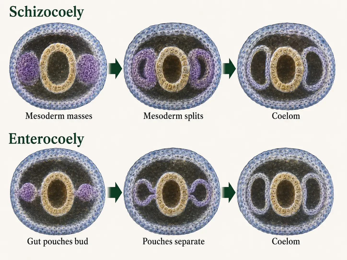
Figure 19. Coelom Development. In schizocoely, coelomic spaces open within paired masses of mesoderm. In enterocoely, pouches of the developing gut bud outward and separate. Both pathways can produce a coelom fully lined by mesoderm, but neither pattern is universal for a major evolutionary branch.
Check Your Understanding Protostome and Deuterostome Developmental Patterns
The Bilaterian Story Continues
Most familiar animals belong to Nephrozoa. One major branch, Protostomia, includes flatworms, rotifers, annelids, mollusks, roundworms, tardigrades, and arthropods. The animals traditionally grouped as Deuterostomia include echinoderms, hemichordates, and chordates, the group that includes vertebrates.
What do these Nephrozoan animals have in common?
These animals share the basic bilaterian body plan introduced in this chapter. They develop from three germ layers, have a clear anterior-to-posterior axis, and inherited a body plan that was ancestrally bilateral.
Why are their bodies so different?
After these lineages split from one another, each followed its own evolutionary path. Different groups evolved new ways to move, feed, grow, sense the environment, support the body, and reproduce. These changes produced the enormous variety of bilaterian animals we see today.
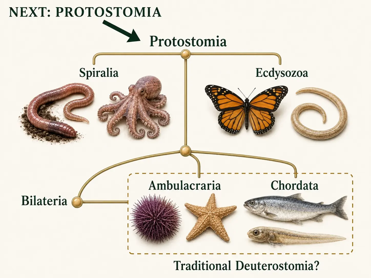
Figure 20. The Bilaterian Story Continues. Earthworms and octopuses belong to Spiralia, while roundworms and butterflies belong to Ecdysozoa. Together, these lineages form Protostomia.
Before You Continue
Complete these activities to finish the Introduction to Animal Diversity module.
-
Watch the Lecture
Watch the lecture
Reinforce the chapter’s major concepts with the Introduction to Animal Diversity lecture.
-
Open the Review
Work through the chapter review
Explore six concept areas through practice questions, explanations, and links back to the chapter. Use the feedback to identify concepts that need another look.
-
Open the Study Guide
Complete the study guide
Recall vocabulary, comparisons, and evolutionary patterns from memory. Compare your responses with the essential points, then revisit cards marked Not Mastered.
-
Open the Lab
Begin the animal lab
Lecture + Lab Courses
If you are enrolled in a lecture-and-lab course, complete the first part of The Origin and Evolution of Animals lab. Stop before Exercise G: Protostomes Overview.
-
Complete the Canvas quiz
Required Course Task
Return to your course in Canvas and complete the Introduction to Animals quiz to finish this module.
© 2026. Jason Walker, PhD. All rights reserved.