Opisthokonta and the Origin of Fungi
Along with animals and many unicellular eukaryotes, fungi make up the supergroup Opisthokonta within Eukarya. All unicellular organisms within this group have posterior flagellate cells used in propulsion. While this trait is uncommon among fungi and animal cells, remnants of the posteriorly flagellated cells can still be seen in animal sperm cells and among the motile gametes and spores of chytrid fungi.
Opisthokonta is divided into two main groups: Holozoa (animals and their relatives) and Holomycota (fungi and their relatives) . Within both of these groups are several examples of unicellular species, and it is believed that multicellularity evolved independently in both fungi and animals.
All members of the Holozoa take food internally and absorb the nutrients once in the body (internal digestion). Animals (Kingdom Animalia) are all multicellular organisms with internal digestion. Animals are most closely related to unicellular or colonial choanoflagellates (Phylum Choanozoa), and are thought to have originated from organisms similar to modern-day choanoflagellates. Sponges (Phylum Porifera) have tissues with cells that strongly resemble choanoflagellates.
All members of Holomycota absorb their food with external structures (external digestion). For example, certain fungi decompose wood with cells known as hyphae that excrete digestive enzymes into the wood cells and absorb the nutrients back into the body of the fungus. Fungi (Kingdom Fungi) are most closely related to another group of unicellular organisms (Order Nucleariida). Nucleariids are amoeba-like organisms with filose (thread-like) pseudopodia. These pseudopodia assist the nucleariid with cell movement as well as sensing and consuming prey.
While fungi can be multicellular or unicellular, all fungi have two things in common. First, they all absorb their food externally. Second, all fungi have cell walls made of a tough polysaccharide, called chitin. This allows the fruiting bodies of fungi (i.e. mushrooms) to have upright growth. Many animals also have chitin. It is what forms human hair and fingernails, and the exoskeleton of insects and crustaceans.
The main difference between fungi and their closest relative (nucleariids) is that all fungi have a cell wall made of the polysaccharide chitin, whereas nucleariids do not. This indicates that chitin likely evolved only once within the common ancestor of both Holozoa and the Holomycota, but was likely lost by an ancestral nucleariid, resulting in no modern nucleariids with cell walls composed of chitin.
While these two characteristics are common among all fungi, the actual relationships among fungal groups are hotly debated, and will likely be majorly altered in the coming years. With that said, there are five groups of fungi that are morphologically similar to each other, even though some of these morphological similarities are not currently supported by genetic analyses.
Diversity of Fungi
Scanning electron micrograph of a microsporidian spore with an extruded polar tubule inserted into a eukaryotic cell. The spore injects the infective sporoplasms through its polar tubule.
Microsporidia
Microsporidia (Division Microsporidia) are unicellular organisms that were once thought to be protists not closely related to fungi. However, genetic analyses have consistently placed species within this division in the fungi kingdom. Upon closer inspection, microsporidia do share certain common characteristics with all fungi. For example, they absorb nutrients and have chitin cell walls. Microsporidia are intracellular parasites of animal cells that absorb nutrients with specialized structures known as polar tubes. These polar tubes penetrate the cell walls of animal cells and absorb nutrients from the animal cell into their bodies.
Chytridomycota
Chytrid fungi (Division Chytridomycota – currently disputed) are predominantly aquatic (or semi-aquatic) fungi that can be unicellular or multicellular. All chytrids have motile cells that move by a single flagellum: gametes (haploid) and spores (diploid). No other fungi have motile spores. While all chytrids share these morphological characteristics, phylogenetic analyses indicate that these organisms make up several different groups, not just one Chytridomycota. In other words, chytrid fungi are not monophyletic, but paraphyletic. This group will most likely be split into several groups.
In the past several decades, many species of amphibians have been dying off in staggering numbers across the entire planet. While many factors have been proposed, it appears the culprit is a species of chytrid that dries the amphibians’ skin. This drying essentially suffocates the amphibians as they breathe directly across their skin.
Some chytrids are parasites on algae. The big structure in picture 1 and 2 is a unicellular alga and the little small sphere is the zoosporangium (fruiting body of the chytrid fungus. When the zoosporangium opens, it releases zoospores. Chytrid zoospores are unique as they are the only ones that have a whip-like flagellum. They use this flagellum to swim in water in attempt to find a host. Once they find a host, the flagellum retracts and the chytrid will begin to parasitize its new host. eventually developing a new zoosporangium.
Zygomycota
Zygomycetes (Division Zygomycota – currently disputed) are terrestrial, multicellular fungi that produce hundreds of non-motile spores in an extremely resilient, thick walled sporangium. Reproduction is most commonly asexual in globular sporangia, held upright by hyphae. However, sexual reproduction occurs when two sporangia fuse, creating a zygosporangium, allowing fertilization of the different mating organisms’ haploid gametes, creating a diploid spore. A common example of a zygomycete is black bread mold (Rhizopus stolonifer). While all zygomycetes share these morphological characteristics, the phylogenetic relationships of this group are very unclear. Current analyses suggest that there are actually several different clades of species that are considered zygomycetes, suggesting that zygomycetes are also paraphyletic, not monophyletic.
Glomeromycota
Glomeromycetes (Division Glomeromycota) form arbuscular mycorrhizas (AMs) existing in a symbiotic relationship within the roots of land plants. Fungal hyphae are only one cell thick, whereas plant cells are several cells thick. Therefore, fungal hyphae are able to penetrate far more completely into the soil, absorbing water and nutrients far more efficiently than plants can. The hyphae also penetrate into the root of the plant, allowing the plant to absorb the water and nutrients from the fungus. In return, the plant provides the fungus sugars produced via photosynthesis. This relationship is so efficient that glomeromycetes are present in the majority (>80%) of all plant species analyzed.
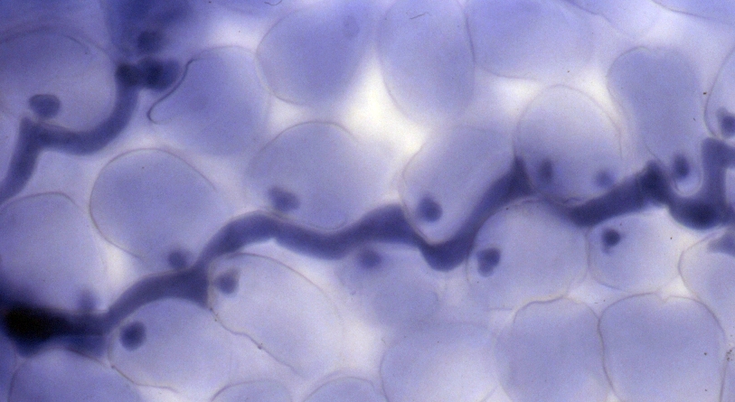
Glomeromycetes are arbuscular mychorrhizal fungi, which exist in symbiotic relationships with most plant species. In this photo, the large stick-like structures are the roots of a plant. The thin, white filaments are the hyphae. The reproductive structures of the fungi are the orange-brown globular structures.
Ascomycota
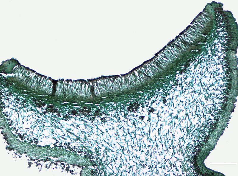
Ascomycetes (Division Ascomycota) are known as “sac fungi,” referring to their defining characteristic, the ascus. The ascus is a sexual structure in which non-motile spores are held. Unlike zygosporangia, which hold hundreds of spores, each ascus will hold between 5 and 12 spores. Asci are typically aggregated together in a structure known as the apothecium. Ascomycota is a monophyletic group within the subkingdom Dikarya, which includes the basidiomycetes. All members of Dikarya have the unusual characteristic of harboring two nuclei.
Over half of ascomycetes are involved in a symbiotic relationship with photosynthetic cyanobacteria or algae, in structures known as lichens. The fungus protects the photosynthetic cells in a tissue known as the thallus, anchoring to a substrate, and absorb water and nutrients. The cyanobacteria or algal cells provide the fungus carbohydrates from photosynthesis. This symbiotic relationship allows lichens to thrive in virtually inhospitable environments, such as directly on rock.
Basidiomycota
Basidiomycetes (Division Basidiomycota) are known as “club fungi,” referring to their basidia, club-shaped pedestals on which non-motile spores are held, usually in fours. Most common mushrooms are basidiomycetes, but this group also includes puffballs, shelf fungi, chanterelles, smuts and rusts. Nearly all basidiomycetes are multicellular (yeasts are the exception), feeding via an extensive network of hyphae and reproducing via basidiospores, those spores held up on a basidium. Most basidiomycetes are saprophytic, obtaining nutrients from decomposing organic matter. Basidiomycetes are the only multicellular organisms capable of digesting the tough polysaccharide that makes wood cells, lignin.


