The Origin and Evolution of Animals*
*This webpage is in draft form. It is incomplete. You will need to complete some outside research to complete your lab. I apologize for any inconvenience.
Animals (Kingdom Animalia) are all multicellular organisms that internally digest their food. Many fungi are also multicellular; however fungi differ from animals by digesting their food externally and absorbing the digested food into their body. Both of these groups belong to the Eukarya supergroup, Opisthokonta.
Phylum Choanozoa
Not all organisms in Opisthokonta are multicellular. Phylogenetic analyses have indicated that animals closest living relative is the unicellular choanoflagellate (Phylum Choanozoa). Choanoflagellates represent a transitionary model from protist (single celled eukaryote) to animal. They are ovoid-shaped cells easily identified by the presence of a collar and a long, single flagellum (Fig. 1). They eat by entrapping bacteria and detritus into the collar by moving its flagellum and then engulfing the prey via endocytosis. In this manner, choanoflagellates are similar to animals in that they digest their food internally. Some species of choanoflagellates form colonies (Fig. 2). Grouping together increases each cell’s chance of capturing food. Colonies are unicellular organisms that are physically connected to each other, but do not have any differentiation of tissues. Individual cells can break free from the colony and continue fully functioning in its absence.

Figure 1. A single, unicellular choanoflagellate. Choanoflagellates are easily identified by the collared, ovoid cell win a single, long flagellum.

Figure 2 A colony of choanoflagellates. Interestingly, some species of choanoflagellates can form simple colonies.
Phylum Porifiera
All animals are multicellular, meaning that they have different types of cells. The simplest animals living today that represent multicellularity are commonly known as sponges (Phylum Porifera). Sponges are filter feeders in marine environments that have very porous bodies, allowing for the constant circulation of nutrient-containing water. Interestingly, the feeding cells of sponges (known as choanocytes, Fig. 3) closely resemble the morphology of choanoflagellates (Fig. 1). These specialized cells have flagella that trap food particles within them (similar to choanoflagellates) and either digest the particles or shuttle the food to other cells. This differentiation of cell types makes sponges multicellular. Unlike sponges, choanoflagellate cells do not share food particles. While members of Porifera have a few different cells, those cells are not separated from each other by membranes. In this regard, it is said that sponges do not have “true” tissues. All other animals have true tissues. Sponges are radically different from all other animals. They have an asymmetrical body plan (Fig. 4) and lack nerves and muscles, whereas all other animals have symmetry and contain nerves and muscles.
_(14763631314).jpg)
Figure 3. Cross section of a sponge. The digestive layer of sponges contain choanocytes, which capture food particles with their flagella and entrap the food in the collars of the cells.

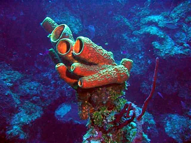

Figure 4. Asymmetry. Sponges also differ from all other animals in that they have an asymmetrical body plan and do not have nerves, muscles, a digestive or a circulatory system. Radial Symmetry. Cnidarians (i.e. jelly fish) and Ctenophorans (box jellies) have radial symmetry, consisting of many planes of symmetry. Bilateral Symmetry. Most animals are bilaterally symmetrical, consisting of two symmetrical halves.
Diploblasty
The differentiation of “true” tissues in the animal kingdom set the stage for the a major radiation of diversification in the animal kingdom. The earliest animals with true differentiation of tissues are known as diploblasts. This refers to the emergence of two tissue layers during the development of the embryo: the ectoderm and the endoderm (Fig. 5). The ectoderm becomes the outer layer (skin) of the animal, where the endoderm develops into the digestive system (see Fig. 6). Diploblastic organisms are all radially symmetrical (Fig. 4) and have a digestive and circulatory system. Diploblasts also have a nerve net, a system of nerve cells attached to each other in a regular network (Fig. 8). Unlike other animals, diploblasts do not have a concentration of nerves, as is the case of the human brain. Diploblasts are comprised of two phyla: Cnidaria (including jellyfish, anemones and coral) and Ctenophora (box jellies).

Figure 6. A comparison of diploblasty vs. triploblasty of the blastula. During early embryonic development in animals, undifferentiated cells begin to differentiate. In diplobastic animals the initial differentiation produces two distinct germ layers: the endoderm and ectoderm. In all animal, these layers develop into the digestive system and outer layer (i.e. skin), respectively.

Figure 7. Gastrulation of a diploblast. The formation of germ layers from a (1) blastula to a (2) gastrula. Certain ectoderm cells (orange) move inward forming the endoderm (red). The pore created during gastrulation, known as the blastopore, becomes the opening responsible for ingestion and excretion.
Cnidaria
Cnidarians (Phylum Cnidaria) are either free-swimming medusas (e.g. jellyfish and hydra) or sessile polyps (e.g. sea anemones and coral). All of these organisms are predators that capture their prey using specialized stinging cells, cnidocytes. All cnidarians are radially symmetrical, which allows these organisms to detect potential prey in all directions. Adult cnidarians have a digestive system that stems from the endoderm of the developing embryo. The ectoderm develops to become the exterior of the organism, which includes muscles, allowing these organisms to move.

Figure 8. Diploblasty in cndiarians (jellyfish-left, anenome-right). The external layer of the adult (red) emerges from the ectoderm, whereas the internal digestive organ emerges from the endoderm.

Figure 9. Example of a nerve net in a hydra. Nerve and muscles originated in the ancestors of Cnidaria and Ctenophora. The nerve net allows these organisms to sense and react to their environment. However, unlike triploblastic animals there is no localized collection of nerves.
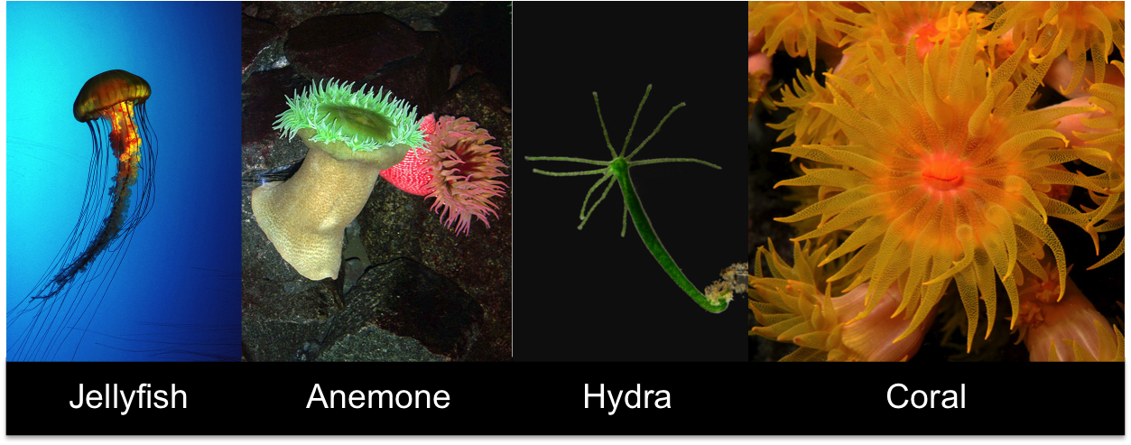
Figure 10. Phylum Cnidaria. Characteristics include diploblasty, a nerve net, cnidocytes and radial symmetry.
Ctenophora
Comb jellies (Phylum Ctenophora) are free-swimming planktonic predators that move by beating cilia in an organized pattern. Like cnidarians, they are diploblastic and contain a nerve net and simple muscles. Some comb jellies have stinging cells (cnidocytes), while the majority capture their prey with sticky cells. Some deep dwelling ctenophores are bioluminescent. A species of box jelly in Australia contains the most potent venom of any animal on the planet. Certain bodies of water are completely forbidden for humans to enter, as contact with this box jelly only a few centimeters in length can kill a person even before they can swim ashore by violently attacking the nervous system and heart.

Figure 11. Phylum Ctenophora. Characteristics include: diploblasty, radial symmetry and cilia.
Triplobasty

Figure 12. Triploblastic animals are characterized by a central nervous system and cephalization.
Other than Porifera, Cnidaria, and Ctenophora, all animals on earth are triploblastic. This refers to the presence of a third germ layer in the development of the blastula: the mesoderm (see Fig. 6 above). While the ectoderm develops into the skin and the endoderm develops into the digestive system, the mesoderm develops into everything else: muscles, bones, and all non-digestive organ systems. All triploblasts share other characteristics. While diploblasts have a non-differentiated nerve net, triploblasts have a centralized nervous system (Fig. 12), a concentration of nerve cells in ganglia and networks known as tracts. This aggregation of neurons has led to the development of cephalization (Fig. 12), a concentration of sensing organs (i.e. eyes, ears, and noses) and feeding structures towards a head region. Cephalization has promoted bilateral symmetry (Fig. 4c) over radial symmetry, with a defined left and right half.
Phylum Acoelomorpha
Members of Phylum Acoelomorpha are the most primitive bilateral triploblastic animal (Fig. 13). They differ from other triploblasts in that they have never ancestrally developed a body cavity, known to biologists as a coelom. Most acoelopmorphates live in mud and sand in marine environments and move by beating cilia.

Figure 13. Phylum Acoelomorpha. Characteristics include a bilateral body plan, cephalization (notice the eyespots on the upper right) and a centralized nervous system. However, unlike all other bilateral animals acoelomates lack a coelom.
Coelomates: Protostomes and Deuterostomes

Figure 14. Developmental differences between coelomates: the protostomes and deuterostomes.
The next major evolutionary milestone in animals was the development of a coelom, a fluid-filled body cavity (Fig. 14). These organisms are known as coelomates, and can be divided into two main groups based on how the embryo develops: protostomes and deuterostomes.
As a coelomate’s fertilized embryo begins to transform from a group of undifferentiated cells (a blastula), an indention forms, known as a blastopore (Fig. 14). The blastopore continues to grow exiting out the opposite end of the developing embryo and forming a tube that becomes the animal’s digestive system. In protostomes, the blastopore becomes the mouth; while in the deuterostomes the blastopore becomes the anus. As the blastopore elongates in the developing embryo, the coelom begins to develop in two different ways in protostomes and deuterostomes. In protostomes, the coelom develops by blocks of mesoderm hollowing out. In deuterostomes, the coelom develops from pockets of mesoderm near the endoderm pinching off to eventually becoming a coelom. While the development of the coelom varies for protostomes and deuterostomes, the end result is the same: a simple spherical embryo with a digestive system composed on one end with a mouth and the other with an anus. In deuterostomes blastula divides in a pattern known as radial cleavage, with cellular division occurring parallel or perpendicular to the major polar axis. In protostomes the cleavage is called spiral because division planes are oriented obliquely to the polar major axis.
Protostomes: Lophotrochozoa and Ecdysozoa
Protostomes are split into two main groups: the Lophotrochozoa and the Ecdysozoa. While members within these groups can be morphologically diverse, the way members of each group grow is similar. Ecdysozoa all shed their exoskeleton in order to expand their bodies (Fig. 15). This process is known as molting. For example, insects are trapped inside a fixed body due to their rigidity of their exoskeleton. In order to grow in size, insects have to leave their exoskeleton behind. Once they molt, they expand their bodies and form another, bigger exoskeleton. Lophotrochozoa all grow incrementally by extending their skeleton (Fig. 16). For example, clams add layers to their shell and stretch their bodies to grow.

Figure 15. An ecdysozoan molting. Ecdysozoans must shed their outer exoskeleton in order to increase in size.

Figure 16. Incremental growth occurs in lophotrochozoans. The radiating rings of a clams shell is caused by varying minerals in the shifting sands as a clam develops.
Phylum Bryozoa (Protostomes: Lophotrochozoa)
Lophotrochozoa are a widely variable group of coelomate triploblastic animals. One of the earliest Lophotrochozoans to appear in the fossil record during the Cambrian Explosion are known as moss animals or bryozoans (Phylum Bryozoa). Bryozoans are typically colonial organisms that appear more closely related to a sponge than a bilateral organism. However, on closer inspection, the individual organisms are bilateral. Bryozoans feed with a special structure known as a lophophore, a ring of ciliated tentacles surrounding a mouth. These cilia beat towards the mouth, effectively drawing food particles towards the mouth.
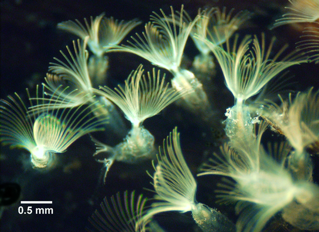
Figure 17. Phylum Bryozoa. Moss animals represent one of the earliest groups of lophotrochozoans.
Phylum Rotifera (Protostomes: Lophotrochozoa)
Rotifers (Phylum Rotifera) are very small (<1cm) aquatic filter feeders that use a special grouping of cilia, known as a corona, surrounding the mouth which reigns in food particles. The corona resemble the lophophore of the Bryozoans, but are highly reduced. While a few rotifers are sessile, most are able to move with use of their corona.

Figure 18. Phylum Rotifera. Rotifers are small animals that feed with specialized mouthparts known as a corona.
Phylum Platyhelminthes (Protostomes: Lophotrochozoa)
Flatworms (Phylum Platyhelminthes) are broad, flattened worms that were once thought to be closely related to Acoelomorpha due to the fact that they are bilateral, triploblastic organisms that do not have a coelom. However, phylogenetic analyses have consistently placed this group within the Lophotrochozoa. This indicates that these organisms have evolutionarily lost their coelom. There are three groups of flatworms: turbellarians, cestodes, and trematodes.
Turbelllarians (Phylum Platyhelminthes)
Turbellarians are primarily free-living aquatic predators of small organisms. Planarians are some of the best known examples of turbellarians. Some planarians have the unique ability of being able to regenerate lost body parts.

Figure 19. Cestode in Phylum Platyhelminthes.

Figure 20. Trematode in Phylum Platyhelminthes.
Cestodes (Phylum Platyhelminthes)
Cestodes are internal parasites completely dependent upon their host for nutrients. They are so dependent that many of them have evolutionarily lost a digestive system. The best known example is a tape worm. Characteristics of all Platyhelminthes include an evolutionary loss of coelom, yet bilaterally symmetrical and triploblastic. Cestodes are internal parasites of animals that have evolutionarily lost their mouth parts and digestive system, absorbing digestive food from their host directly.
Trematodes (Phylum Platyhelminthes)
Trematodes are also internal parasites of animals. But unlike cestodes, trematodes retain mouthparts and a digestive system. Humans can be afflicted with trematodes known as liver flukes.
Phylum Annelida (Protostomes: Lophotrochozoa)
Unlike flatworms, segmented worms (Phylum Annelida) have true coeloms with a circular body separated by segments. Each of these segments are practically identical in their external and internal composition of organs. Only the front segments and the rear segments have specialized organs. Front segments contain a mouth, crop, gizzard and primitive hearts called aortic arches. There are three types of annelids.
Polychaetae (Phylum Annelida) - Polychaete worms are segmented worms that have muscular extensions (parapodia) extending from their segments, in which bristle-like hairs (chaetae) are attached.
Oligochaetae (Phylum Annelida) - Oligochaete worms have lost their parapodia. They still retain some chaetae. However, they have far fewer chaetae per segment than polychaetes. Earthworms are oligochaetes.
Hirundinea (Phylum Annelida) - Hirundinea have completely lost both parapodia and chaetae. Leeches are an example of Hirundinea and are an ectoparasite of many mammals, including humans.
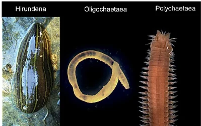
Figure 21. Phylum Annelida. Characteristics includes triploblasty, bilateral symmetry, cephalization, presence of a coleom and segmentation.
Phylum Mollusca (Protostomes: Lophotrochozoa)
Molluscs (Phylum Mollusca) are a widely variable group of organisms that have two defining characteristics. All molluscs have a mantle, a single internal cavity used for breathing and digestion. Some molluscs (i.e. clams, snails, and nautiluses) secrete calcium carbonate from their mantles, creating protective shells. All molluscs have some version of a muscular foot.
Gastropoda (Phylum Mollusca)
Gastropods (Fig. 23) include snails and slugs. These organisms have a large muscular foot on their underside which is primarily responsible for their movement. Snails have a single, spiralling shell. Slugs have evolutionarily lost their shells. Gastropods are typically herbivores that feed by a special scraping mouthpart known as a radula.
Bivalvia (Phylum Mollusca)
Bivalves (Fig. 23) are molluscs that have two shells that are able to open and close on a hinge. Most bivalves are suspension feeders, eating any small animal or protists that filter into the mantle and are collected via gills. Bivalves are the only mollusc group that do not feed via radula.

Figure 22. Subphylum Gastropoda. Like all mollucs, snails have a muscular foot and a mantle.

Figure 23. Subphylum Bivalvia.
Polyplacophora (Phylum Mollusca)
Chitons are similar to snails in several ways. They move with a muscular foot and ingest food (typically vegetation) with a radula, a scraping mouthpart. Chitons are uniquely different from snails in that they have a flattened body with eight shell plates, where snails have a continuous coiling shell.
Cephalopoda (Phylum Mollusca)
Cephalopods include octopuses, squid and nautiluses. Their muscular foot has adapted by separating into tentacles. Of this group, only nautilus retain a hard outer shell. Cephalopods represent the most intelligent invertebrates in the animal world.