Chapter: Meiosis
Introduction to Meiosis
Meiosis is a form of nuclear division that is similar to mitosis in some ways, but it produces a very different outcome. While mitosis creates daughter cells that are genetically identical to the parent cell and maintain the same chromosome number, meiosis produces cells containing only half of the original genetic material.
The cells produced by meiosis are called gametes, which include sperm cells and egg cells. These specialized reproductive cells are required for sexual reproduction. Starting from a single diploid cell, meiosis produces four daughter cells. Each daughter cell contains half the DNA of the original parent cell. Although the chromosome number is reduced, the daughter cells are not genetically identical. Instead, each gamete contains a unique combination of chromosomes produced through processes such as crossing over and independent assortment.
This genetic variation is extremely important because it ensures that no two gametes are genetically identical. During sexual reproduction, gametes from two organisms fuse during fertilization. This restores the full chromosome number and produces genetically unique offspring. The continual reshuffling of genetic material increases variation within a population, strengthening the gene pool and improving a species’ ability to adapt to changing environmental conditions over time.
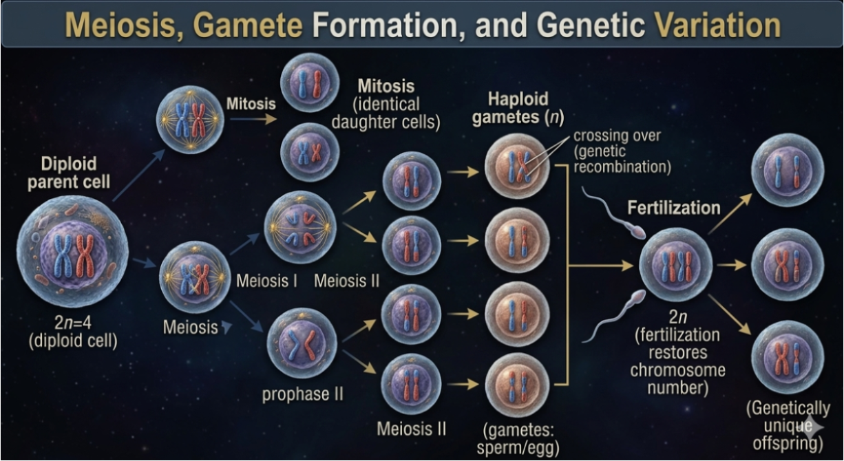
Figure 1. Meiosis. Meiosis produces four genetically unique haploid gametes through two sequential divisions and genetic recombination, while fertilization restores diploidy and generates genetically diverse offspring, increasing variation within populations and improving adaptive potential over time.
Chromosomes and Sexual Reproduction in Humans
Humans have 46 chromosomes in total. Of these, 23 chromosomes are inherited from the mother and 23 chromosomes are inherited from the father.
During fertilization, each chromosome from one parent pairs with a corresponding chromosome from the other parent. These matching chromosome pairs are called homologous chromosomes. Homologous chromosomes carry genes for the same traits and contain the same types of genetic information, although the specific versions of those genes (alleles) may differ between the maternal and paternal chromosomes.
When fertilization occurs, the maternal and paternal chromosome sets combine to form a diploid cell. The term diploid means that the cell contains two complete sets of chromosomes, expressed as 2n = 46. In humans, the diploid number is 46 chromosomes. As a result, most cells in the human body contain 46 chromosomes arranged into 23 homologous pairs. Each homologous pair consists of one maternal chromosome and one paternal chromosome carrying genes for the same biological traits.
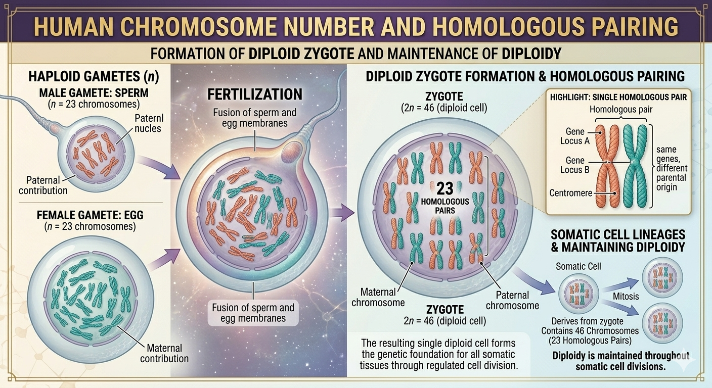
Figure 2. Humans and Meiosis. Meiosis produces four genetically unique haploid gametes through two sequential divisions and genetic recombination, while fertilization restores diploidy and generates genetically diverse offspring, increasing variation within populations and improving adaptive potential over time.
Interphase Before Meiosis
Before meiosis begins, the cell passes through interphase, which occurs in the same general manner as interphase before mitosis. During this stage, the cell grows, carries out normal metabolic functions, and replicates its DNA in preparation for cell division.
Phases of Interphase: G1, S, and G2
During the G1 phase, each homologous chromosome pair consists of two unreplicated chromosomes: one maternal chromosome and one paternal chromosome of the same type. At this stage, the DNA is relaxed, known as chromatin, and unreplicated forming a solitary double helix.
During the S phase, DNA synthesis occurs and each chromosome is replicated. The replicated chromosome connects two genetically, identical sister chromatids joined together at the centromere. These two sister chromatids are exact copies of one another and remain physically connected, forming a single replicated chromosome (two sister chromatids = one replicated chromosome).
During the G2 phase, each homologous chromosome pair consists of two replicated chromosomes: one maternal chromosome and one paternal chromosome of the same type. At this stage, the chromosomes have been replicated.
Chromosome Organization After Replication
After replication, each homologous pair consists of two replicated chromosomes: one maternal chromosome and one paternal chromosome. Each replicated chromosome is composed of two sister chromatids. Even though a replicated chromosome contains two chromatids, it is still considered a single chromosome because the chromatids share one centromere. Chromatids belonging to other homologous chromosome (maternal versus paternal) are called non-sister chromatids.

Figure 3. Interphase. During interphase, DNA replication in the S phase duplicates each chromosome to form sister chromatids joined at a centromere. After replication, each homologous chromosome pair consists of two replicated chromosomes—one maternal and one paternal—each made of identical sister chromatids, establishing the chromosomal structure required for meiosis.
Steps of Meiosis: Overview
Meiosis I
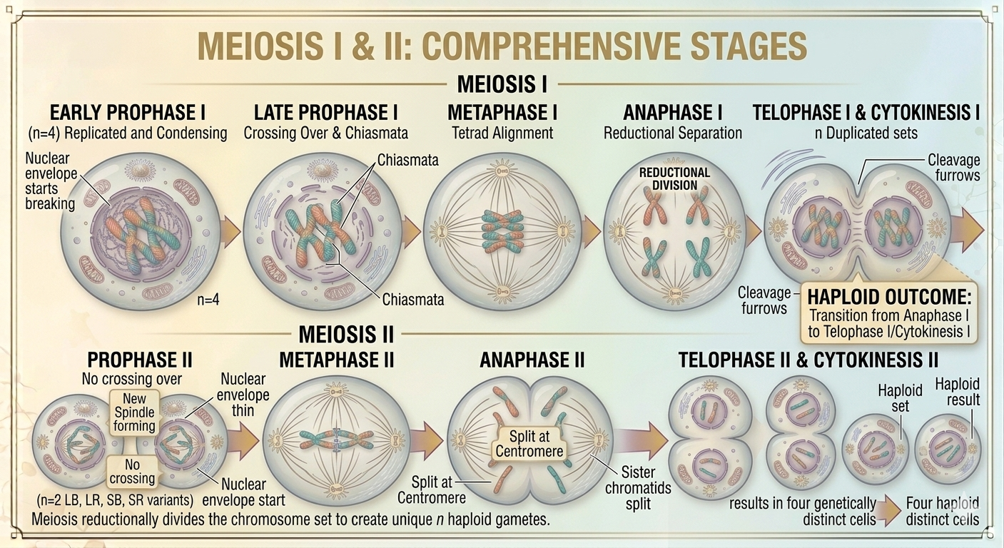
Meiosis I is the first division of meiosis and is responsible for reducing the chromosome number in half from diploid to haploid.
It begins with Prophase I, during which homologous chromosomes pair up in a process called synapsis and exchange genetic material through crossing over, increasing genetic variation. In Metaphase I, homologous pairs (tetrads) align along the metaphase plate, and spindle fibers attach to each homolog from opposite poles. During Anaphase I, homologous chromosomes are pulled apart to opposite poles of the cell, while sister chromatids remain attached. In Telophase I, chromosomes reach the poles and Cytokinesis I divides the cell into two haploid cells.
Meiosis II
Meiosis II resembles mitosis but occurs in haploid cells and does not involve DNA replication beforehand.
It begins with Prophase II, where chromosomes condense again and a new spindle forms in each cell. In Metaphase II, chromosomes align individually at the metaphase plate. During Anaphase II, sister chromatids finally separate and move to opposite poles of the cell. In Telophase II, nuclear envelopes reform, chromosomes decondense, and cytokinesis completes the process.
The final result of meiosis is four genetically distinct haploid cells.
Early Prophase I of Meiosis
Early Prophase I of meiosis begins in a manner similar to mitosis. The chromosomes start to condense, becoming shorter, thicker, and increasingly visible under the microscope. At the same time, the nuclear envelope begins to break down, allowing the cellular machinery access to the chromosomes.
Despite these similarities, meiosis differs from mitosis in one major way during this stage. Homologous chromosomes, one inherited from the mother and one inherited from the father, pair together in a highly specific process called synapsis. Each chromosome aligns precisely with its matching homologous partner to form a structure known as a tetrad. A tetrad contains four chromatids in total: two sister chromatids from the maternal chromosome and two sister chromatids from the paternal chromosome. These chromatids align closely along their entire length, allowing direct physical interaction between homologous chromosomes in the next step, Late Prophase I.
Chromatid Relationships
At this stage, it is important to distinguish between different chromatid relationships. Chromatids belonging to the same replicated chromosome are called sister chromatids. Chromatids belonging to different homologous chromosomes, one maternal and one paternal, are called non-sister chromatids. This distinction becomes especially important during Late Prophase I, when genetic exchange occurs between non-sister chromatids through crossing over.
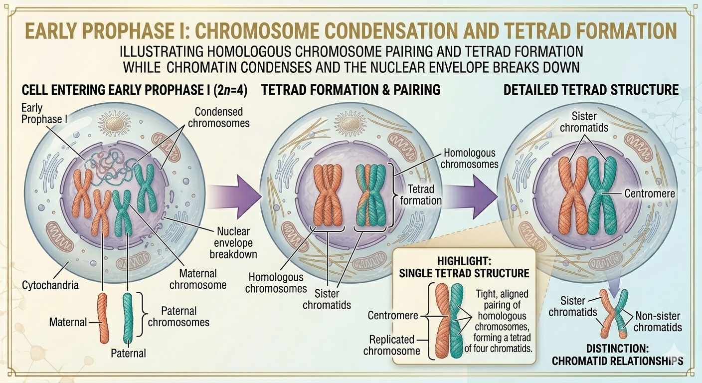
Figure 3. Early Prophase I. During early prophase I, chromosomes condense and the nuclear envelope begins to break down. Homologous chromosomes pair in a highly specific manner to form tetrads, each consisting of four chromatids. This pairing establishes the structural basis for genetic recombination later in meiosis, and distinguishes sister chromatids within chromosomes from non-sister chromatids between homologous pairs.
Late Prophase I of Meiosis
In Late Prophase I, the nuclear envelope disintegrates, and spindle fibers emerge from the centriole pairs. These spindle fibers attach to specialized protein structures called kinetochores, which are located at the centromere regions of each chromosome within the tetrads.
At this point, homologous chromosomes are still paired closely together, but an important genetic process is underway: crossing over. During this process, non-sister chromatids exchange DNA. During crossing over, non-sister chromatids of homologous chromosomes align with remarkable precision so that matching regions of DNA can be exchanged. Enzymes cut the DNA at identical locations on the non-sister chromatids. Then, chromosome segments are swapped and rejoined in a reciprocal manner, causing each chromatid to contain a mixture of maternal and paternal genetic material. The physical points where chromatids remain temporarily connected after this exchange are called chiasmata. These structures are visible evidence that crossing over has occurred and help hold homologous chromosomes together until they separate later in meiosis.
Result of Crossing Over
Crossing over produces recombinant chromatids, which are chromatids containing new combinations of alleles that were not present in either original parental chromosome. This reshuffling of genetic information is a sources of genetic variation in sexually reproducing organisms because genetic material is reorganized at the chromosomal level rather than passed down unchanged.
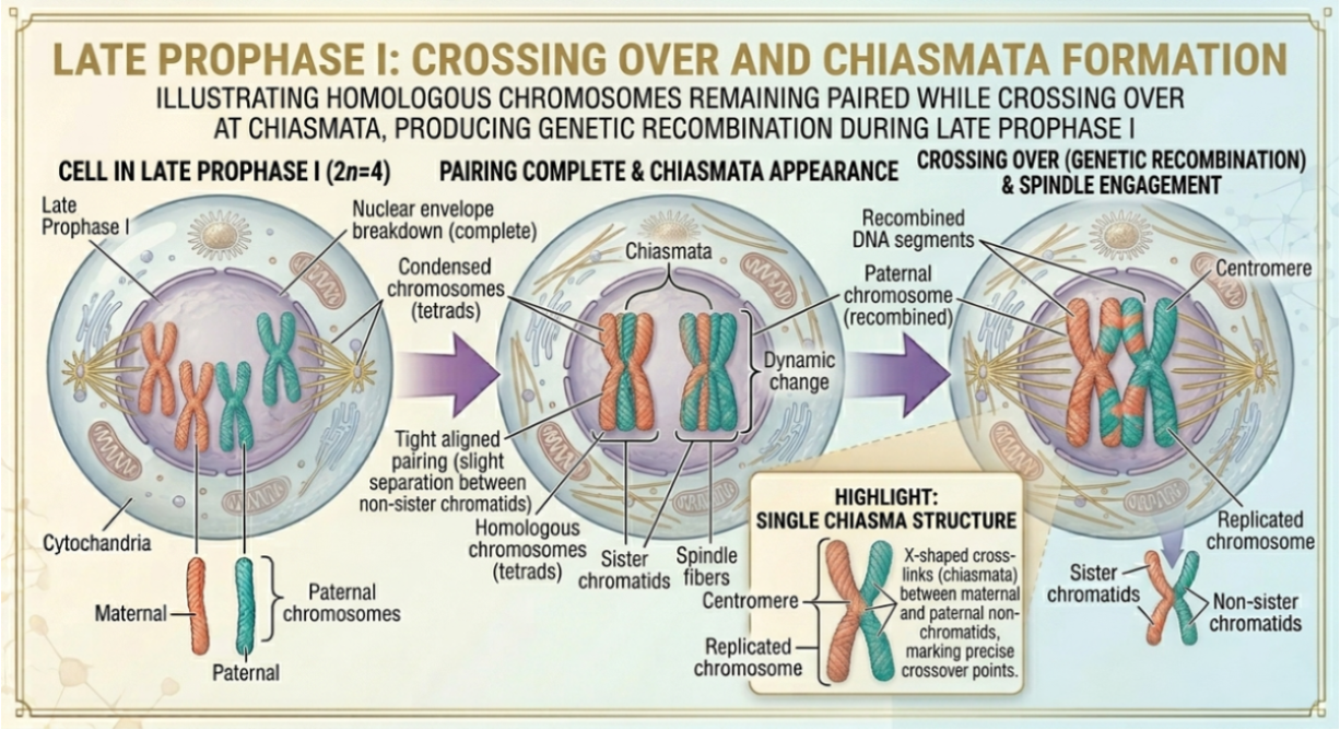
Figure 4. Crossing over during Late Prophase I of Meiosis. During late prophase I, the nuclear envelope fully disintegrates and spindle fibers attach to kinetochores on homologous chromosomes. Tetrads remain paired while non-sister chromatids exchange genetic material at chiasmata through crossing over, producing recombinant chromosomes that increase genetic variation in gametes.
Metaphase I of Meiosis
During Metaphase I, the tetrads align along the metaphase plate, an imaginary plane located at the center of the cell. Each tetrad is positioned so that one homologous chromosome faces one pole of the cell, while the other homologous chromosome faces the opposite pole. By this stage, crossing over is complete. As a result, the recombinant chromatids now contain exchanged segments of maternal and paternal DNA. Spindle fibers attach to the kinetochores of each homologous chromosome within the tetrad. These attachments generate tension that helps properly align the tetrads in the metaphase plate, before separation occurs in the next stage, Anaphase I.
Metaphase I and Independent Assortment
An important feature of Metaphase I is that the orientation of homologous chromosome pairs is random. Maternal and paternal chromosomes can align facing either pole independently of other chromosome pairs. This random alignment contributes to independent assortment, the major source of genetic variation in sexually reproducing organisms. Humans, for example, align 23 tetrads during Metaphase I. Since the homologous pairs align randomly, there are many possible chromosome combinations that can be produced in gametes. In fact there are 2²³, or 8,388,608 possible gamete combinations that one human alone can produce. This means that two humans could produce 2²³ times 2²³ different chromosome combinations, which yields 2⁴⁶ = 70,368,744,177,664. That is over 70 trillion different babies from just two people. And that doesn’t take into account of the genetic variability created by crossing from Late Prophase I.
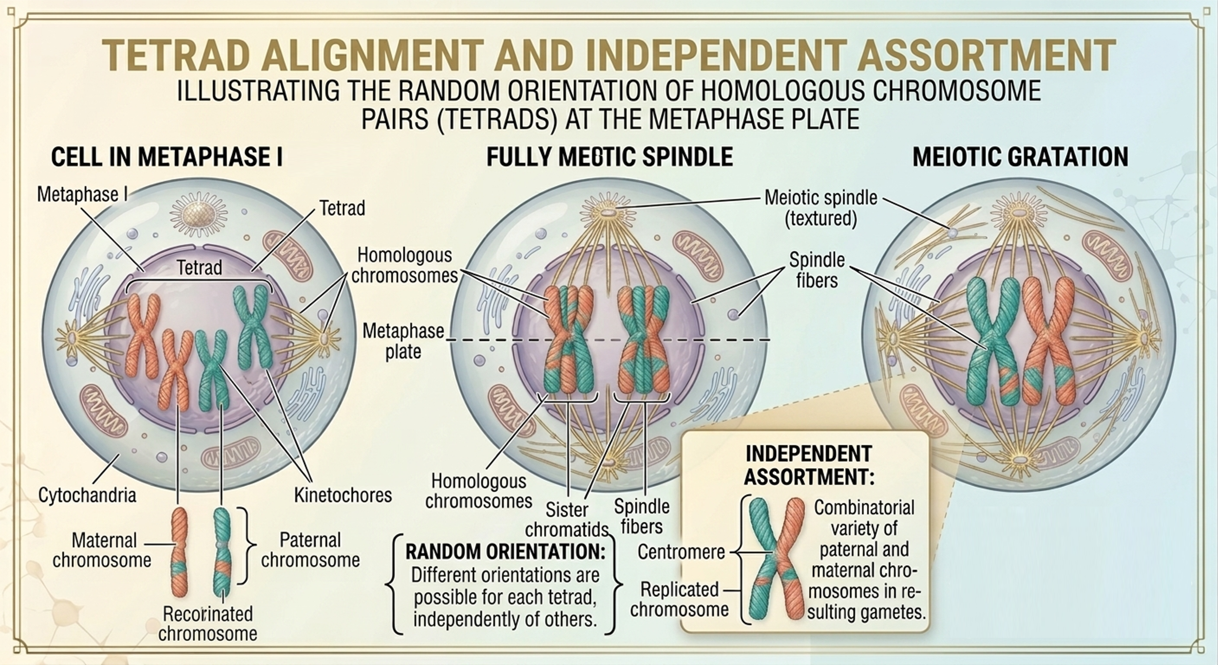
Figure 5. Metaphase I of Meiosis. During Metaphase I, homologous chromosome pairs (tetrads) align along the metaphase plate with spindle fibers attached to opposite kinetochores. The random orientation of each tetrad leads to independent assortment of maternal and paternal chromosomes, generating a large number of possible genetic combinations in gametes.
Anaphase I of Meiosis
In Anaphase I, the homologous chromosomes within each tetrad are pulled apart and move toward opposite poles of the cell. Spindle fibers shorten, generating the force required to separate each homologous pair. A key feature of this stage is that sister chromatids remain attached at their centromeres. Only the homologous chromosomes separate, not the individual chromatids.
As a result of this separation during Anaphase I, each pole of the cell receives a haploid set of chromosomes. However, each chromosome is still in its replicated form and consists of two sister chromatids.
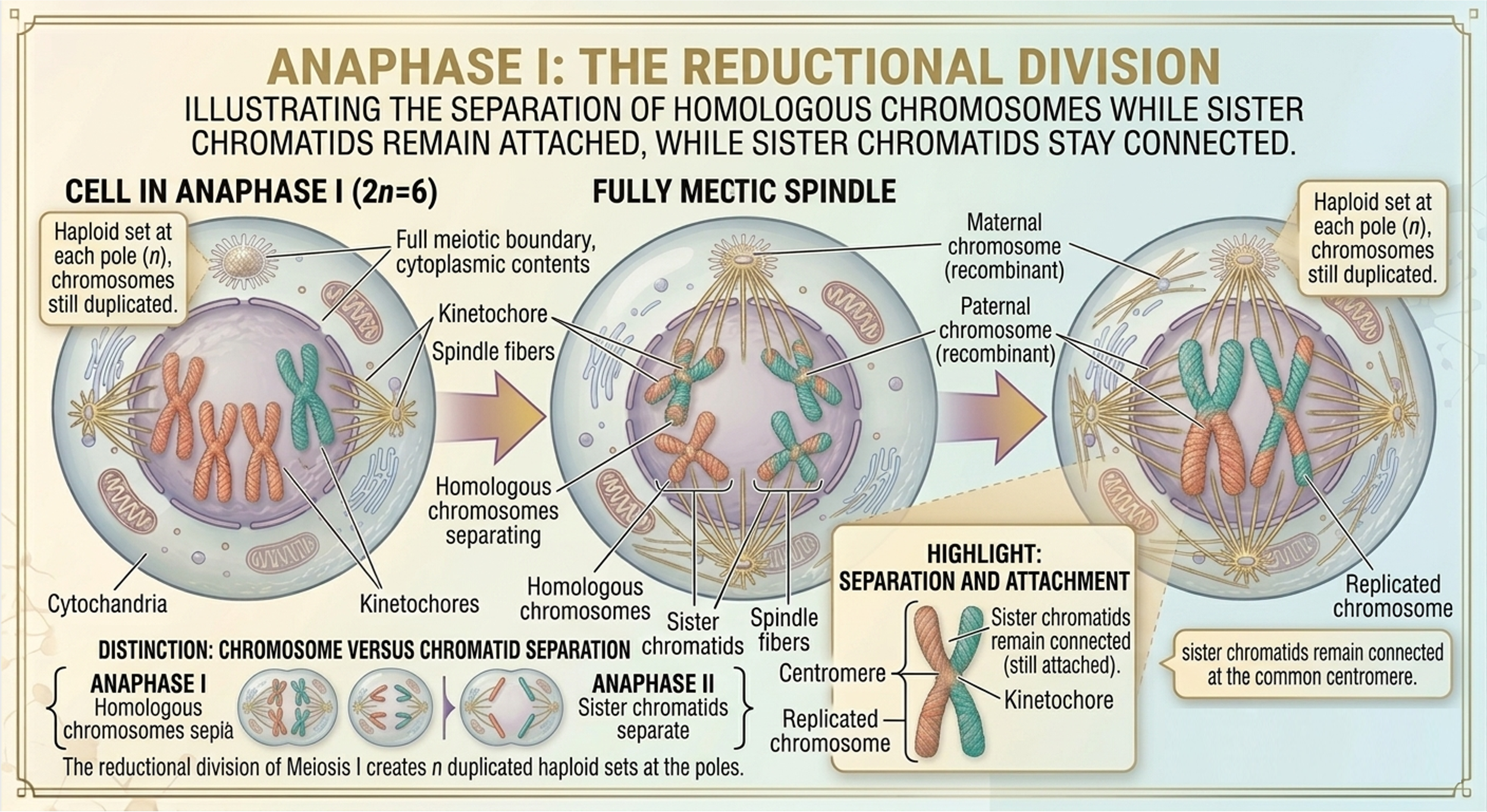
Figure 6. Anaphase I of Meiosis. During Anaphase I, spindle fibers shorten to separate homologous chromosomes and move them toward opposite poles of the cell. Sister chromatids remain attached at their centromeres, meaning only homologous pairs are separated. This reductional division produces haploid sets of recombinant chromosomes at each pole, establishing the chromosome number reduction characteristic of meiosis I.
Telophase I and Cytokinesis I of Meiosis
In Telophase I, the separated replicated homologous chromosomes reach opposite poles of the cell. A nuclear envelope reforms around each set of chromosomes, creating two distinct nuclei. Each newly formed nucleus contains a haploid (n) set of recombinant chromosomes, due to crossing over that occurred earlier in Late Prophase I. Cytokinesis I divides the cytoplasm of the cell. This physical division separates the original cell into two daughter cells. Each daughter cell contains one haploid (n) set of chromosomes.
At the end of Meiosis I, the original diploid cell (2n) has been reduced to two haploid (n) cells. Even though the cell at the end of Meiosis I is considered haploid, the chromosomes are duplicated, meaning that sister chromatids remain attached at their centromeres.
These cells now enter Meiosis II, where the sister chromatids separate to produce four genetically distinct haploid cells.
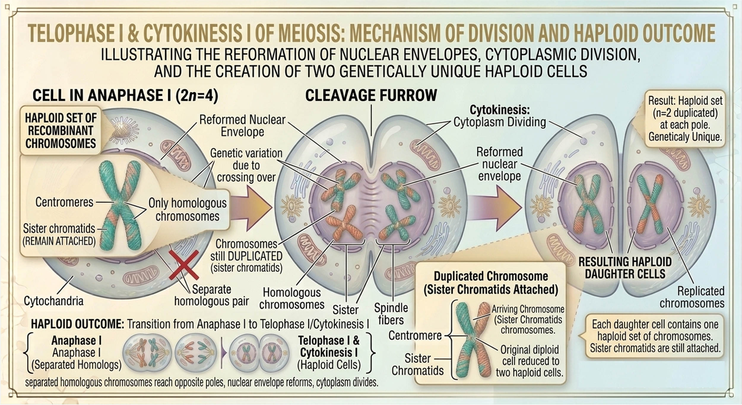
Figure 7. Telophase 1 and Cytokinesis 1. In Telophase I of meiosis, homologous chromosomes reach opposite poles of the cell, and new nuclear envelopes begin to form around each haploid set of chromosomes. Cytokinesis I then divides the cytoplasm, producing two separate haploid daughter cells, each containing chromosomes that are still made of sister chromatids.
Meiosis II Overview
Meiosis II is the second division of meiosis and is responsible for separating sister chromatids. This division ultimately produces four genetically distinct haploid cells.
Steps of Meiosis II: Prophase II, Metaphase II, Anaphase II, Telophase II and Cytokinesis II
In Prophase II, chromosomes condense again if they have relaxed since Meiosis I. The nuclear envelope breaks down if it has reformed, and a new spindle apparatus begins to form in each haploid cell. Centrosomes move toward opposite poles, and spindle fibers attach to the kinetochores of each chromosome, preparing them for alignment. Unlike Meiosis I, there is no pairing of homologous chromosomes and no crossing over in this stage. In Metaphase II, chromosomes line up individually along the metaphase plate. Spindle fibers attach to the kinetochores of each sister chromatid from opposite poles. During Anaphase II, the centromeres split and sister chromatids are pulled apart toward opposite poles, becoming daughter chromosomes. In Telophase II, nuclear envelopes reform around each set of chromosomes as they begin to decondense, marking the near completion of nuclear division. Finally, Cytokinesis II divides the cytoplasm, resulting in four haploid daughter cells.
Result of Meiosis I & II
Meiosis I separates homologous chromosomes in the original diploid (2n) cell, resulting in two haploid (n) daughter cells. Each resulting cell contains a single set of chromosomes, but each chromosome is still replicated and composed of two attached sister chromatids.
Meiosis II separates two replicated, haploid (n) sister chromatids (produced by Meiosis I) into the two, unreplicated haploid (n) daughter chromosomes. From the initial cell that initiated meiosis, four total daughter cells result, each one a haploid cell containing a single set of chromosomes (n) made up of single, unreplicated chromatids. These cells are genetically unique due to crossing over and independent assortment that occurred during Meiosis I.
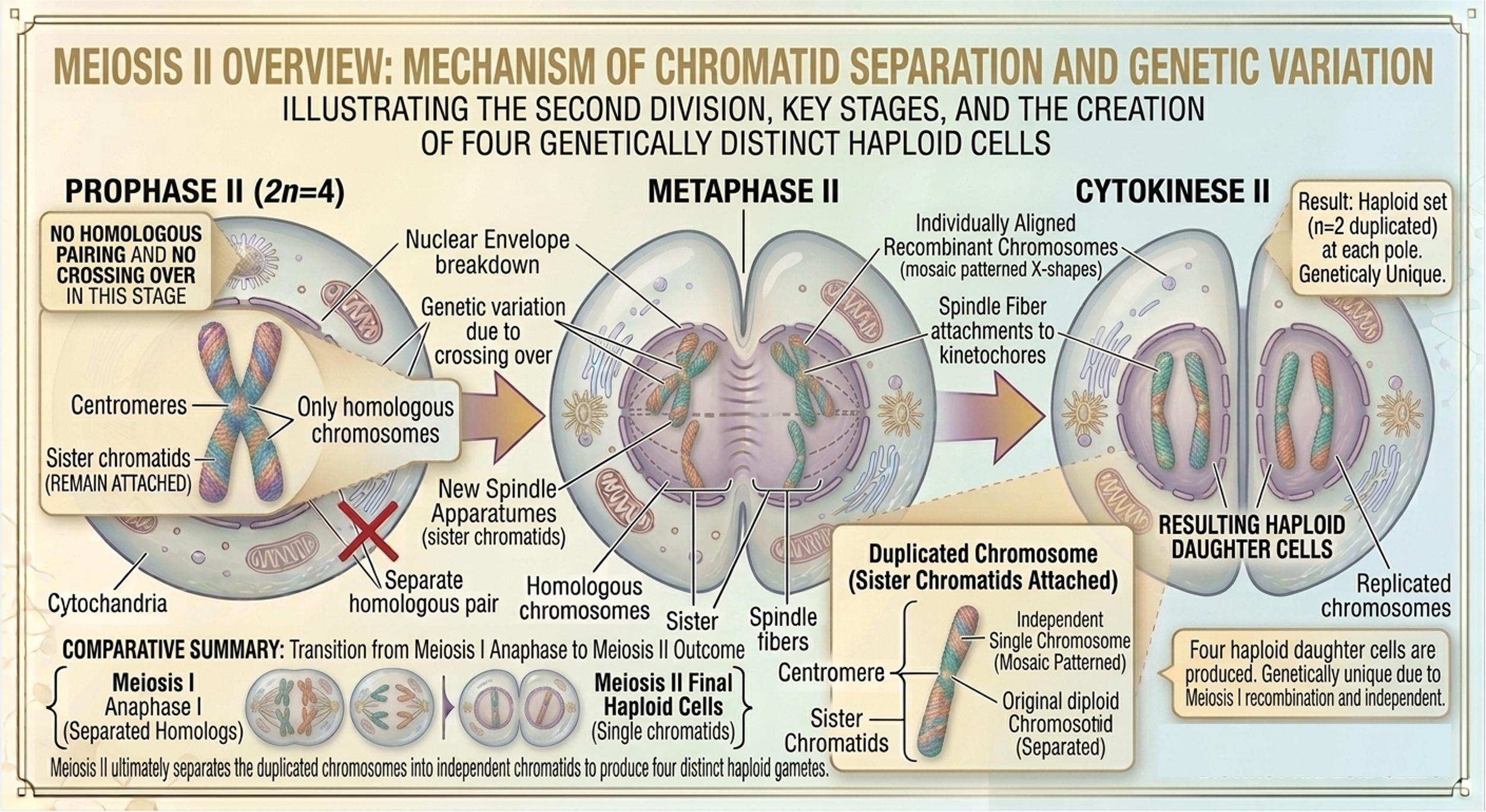
Figure 8. Meiosis II. Meiosis II consists of prophase II, metaphase II, anaphase II, telophase II, and cytokinesis, and it functions to separate sister chromatids without an intervening round of DNA replication. During this division, chromosomes condense and attach to spindle fibers, align at the metaphase plate, separate into individual chromatids, and are enclosed into new nuclei before cytokinesis produces four genetically distinct haploid cells.
Mitosis vs Meiosis
Mitosis and meiosis are both forms of eukaryotic cell division that involve DNA replication, spindle formation, and the segregation of chromosomes to ensure genetic information is passed to daughter cells.
Mitosis
In mitosis, one cell division produces two genetically identical diploid daughter cells. This process is primarily used for growth, tissue repair, and asexual reproduction. During mitosis, chromosomes align individually at the metaphase plate, and sister chromatids separate during anaphase. This ensures that each daughter cell maintains the same chromosome number as the original cell.
Meiosis
In contrast, meiosis consists of two consecutive divisions that produce four genetically distinct haploid cells. These cells are used in sexual reproduction to form gametes. Unlike mitosis, meiosis includes homologous chromosome pairing forming tetrads, as well as crossing over during Prophase I creating recombinant chromosomes. Crossing over and independent assortment increase genetic variation. The first meiotic division separates homologous chromosomes, while the second division separates sister chromatids. As a result, meiosis reduces the chromosome number by half, another form genetic diversity, as the necessary precursors of sexual reproduction.
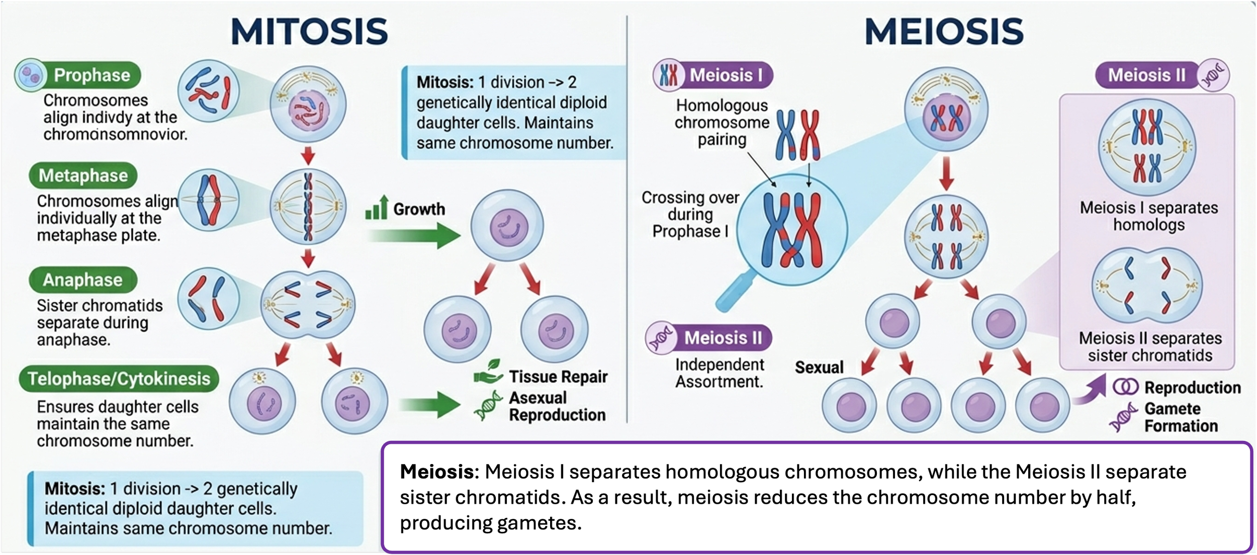
Figure 9. Mitosis vs Meiosis. Mitosis produces two genetically identical diploid daughter cells in a single division, maintaining chromosome number for growth and repair. Meiosis involves two divisions that produce four genetically distinct haploid cells, reducing chromosome number and generating genetic variation through processes like crossing over and independent assortment.